Pharmacological Reviews xPharm- The Comprehensive Pharmacology Reference
HOME HELP FEEDBACK SUBSCRIPTIONS ARCHIVE SEARCH TABLE OF CONTENTS
 QUICK SEARCH:   [advanced]


0031-6997/03/5504-579-581$7.00
Pharmacol Rev 55:579-581, 2003

This Article
Right arrow Abstract Freely available
Right arrow Full Text (PDF)
Right arrow Submit a response
Right arrow Alert me when this article is cited
Right arrow Alert me when eLetters are posted
Right arrow Alert me if a correction is posted
Services
Right arrow Similar articles in this journal
Right arrow Similar articles in PubMed
Right arrow Alert me to new issues of the journal
Right arrow Download to citation manager
Citing Articles
Right arrow Citing Articles via HighWire
Right arrow Citing Articles via Google Scholar
Google Scholar
Right arrow Articles by Catterall, W. A.
Right arrow Articles by Perez-Reyes, E.
Right arrow Search for Related Content
PubMed
Right arrow PubMed Citation
Right arrow Articles by Catterall, W. A.
Right arrow Articles by Perez-Reyes, E.

International Union of Pharmacology. XL. Compendium of Voltage-Gated Ion Channels: Calcium Channels

William A. Catterall, Joerg Striessnig, Terrance P. Snutch and Edward Perez-Reyes

Department of Pharmacology, University of Washington School of Medicine, Seattle, Washington (W.A.C.); Abteilung Pharmakologie und Toxikologie, Institut für Pharmazie, Universität Innsbruck, Innsbruck, Austria (J.S.); Biotechnology Laboratory, University of British Columbia, Vancouver, British Columbia, Canada (T.P.S.); and Department of Pharmacology, University of Virginia, Charlottesville, Virginia (E.P.-R.)

Abstract
Calcium Channel Subunits
Calcium Currents
Calcium Channel Genes
Calcium Channel Molecular Pharmacology

    Abstract
 Top
 Next
 
This summary article presents an overview of the molecular relationships among the voltage-gated calcium channels and a standard nomenclature for them, which is derived from the IUPHAR Compendium of Voltage-Gated Ion Channels.1 The complete Compendium, including data tables for each member of the calcium channel family can be found at http://www.iuphar-db.org/iuphar-ic/.


    Calcium Channel Subunits
 Top
 Previous
 Next
 
Voltage-gated calcium channels mediate calcium influx in response to membrane depolarization and regulate intracellular processes such as contraction, secretion, neurotransmission, and gene expression. Their activity is essential to couple electrical signals in the cell surface to physiological events in cells. They are members of a gene superfamily of transmembrane ion channel proteins that includes voltage-gated potassium and sodium channels (Catterall, 1995Go).

The calcium channels that have been characterized biochemically are complex proteins composed of four or five distinct subunits, which are encoded by multiple genes (Fig. 1) (Catterall, 2000Go). The {alpha}1 subunit of 190 to 250 kDa is the largest subunit, and it incorporates the conduction pore, the voltage sensor and gating apparatus, and the known sites of channel regulation by second messengers, drugs, and toxins. Like the {alpha} subunits of sodium channels, the {alpha}1 subunit of voltage-gated calcium channels is organized in four homologous domains (I–IV) with six transmembrane segments (S1–S6) in each. The S4 segment serves as the voltage sensor. The pore loop between transmembrane segments S5 and S6 in each domain determines ion conductance and selectivity, and changes of only three amino acids (aa) in the pore loops in domains I, III, and IV will convert a sodium channel to calcium selectivity. An intracellular {beta} subunit and a transmembrane, disulfide-linked {alpha}2{delta} subunit complex are components of most types of calcium channels. A {gamma} subunit has also been found in skeletal muscle calcium channels and related subunits are expressed in heart and brain. Although these auxiliary subunits modulate the properties of the channel complex, the pharmacological and electrophysiological diversity of calcium channels arises primarily from the existence of multiple {alpha}1 subunits (Hofmann et al., 1994Go).


View larger version (18K):
[in this window]
[in a new window]
 
FIG. 1. Subunits of CaV1 channels. The subunit composition and structure of calcium channels purified from skeletal muscle are illustrated. The model is updated from the original description of the subunit structure of skeletal muscle calcium channels. This model also fits available biochemical and molecular biological results for other CaV1 channels and for CaV2 channels. P, sites of phosphorylation by cAMP-dependent protein kinase; {Psi}, sites of N-linked glycosylation. Predicted alpha helices are depicted as cylinders. The lengths of lines correspond approximately to the lengths of the polypeptide segments represented.


    Calcium Currents
 Top
 Previous
 Next
 
Calcium currents recorded in different cell types have diverse physiological and pharmacological properties, and an alphabetical nomenclature has evolved for the distinct classes of calcium currents (Tsein et al., 1995). L-type calcium currents require a strong depolarization for activation, are long-lasting, and are blocked by the organic L-type calcium channel antagonists, including dihydropyridines, phenylalkylamines, and benzothiazepines. They are the main calcium currents recorded in muscle and endocrine cells, where they initiate contraction and secretion. N-type, P/Q-type, and R-type calcium currents also require strong depolarization for activation. They are relatively unaffected by L-type calcium channel antagonist drugs but are blocked by specific polypeptide toxins from snail and spider venoms. They are expressed primarily in neurons, where they initiate neurotransmission at most fast synapses and also mediate calcium entry into cell bodies and dendrites. T-type calcium currents are activated by weak depolarization and are transient. They are resistant to both organic antagonists and to the snake and spider toxins used to define the N- and P/Q-type calcium currents. They are expressed in a wide variety of cell types, where they are involved in shaping the action potential and controlling patterns of repetitive firing.


    Calcium Channel Genes
 Top
 Previous
 Next
 
Mammalian {alpha}1 subunits are encoded by at least ten distinct genes. Historically, various names had been given to the corresponding gene products, giving rise to distinct and sometimes confusing nomenclatures. In 1994, a unified but arbitrary nomenclature was adopted in which {alpha}1 subunits were referred to as {alpha}1S for the original skeletal muscle isoform and {alpha}1A through {alpha}1E for those discovered subsequently (Birnbaumer et al., 1994Go). In 2000, a rational nomenclature was adopted based on the well defined potassium channel nomenclature (Chandy and Gutman, 1993Go; Ertel et al., 2000Go). Calcium channels were named using the chemical symbol of the principal permeating ion (Ca) with the principal physiological regulator (voltage) indicated as a subscript (CaV). The numerical identifier corresponds to the CaV channel {alpha}1 subunit gene subfamily (1 to 3 at present) and the order of discovery of the {alpha}1 subunit within that subfamily (1 through m). According to this nomenclature, the CaV1 subfamily (CaV1.1 to CaV1.4) includes channels containing {alpha}1S, {alpha}1C, {alpha}1D, and {alpha}1F, which mediate L-type Ca2+ currents (Table 1). The CaV2 subfamily (CaV2.1 to CaV2.3) includes channels containing {alpha}1A, {alpha}1B, and {alpha}1E, which mediate P/Q-, N-, and R-type Ca2+ currents, respectively (Table 1). The CaV3 subfamily (CaV3.1 to CaV3.3) includes channels containing {alpha}1G, {alpha}1H, and {alpha}1I, which mediate T-type Ca2+ currents.


View this table:
[in this window]
[in a new window]
 
TABLE 1 Physiological function and pharmacology of calcium channels


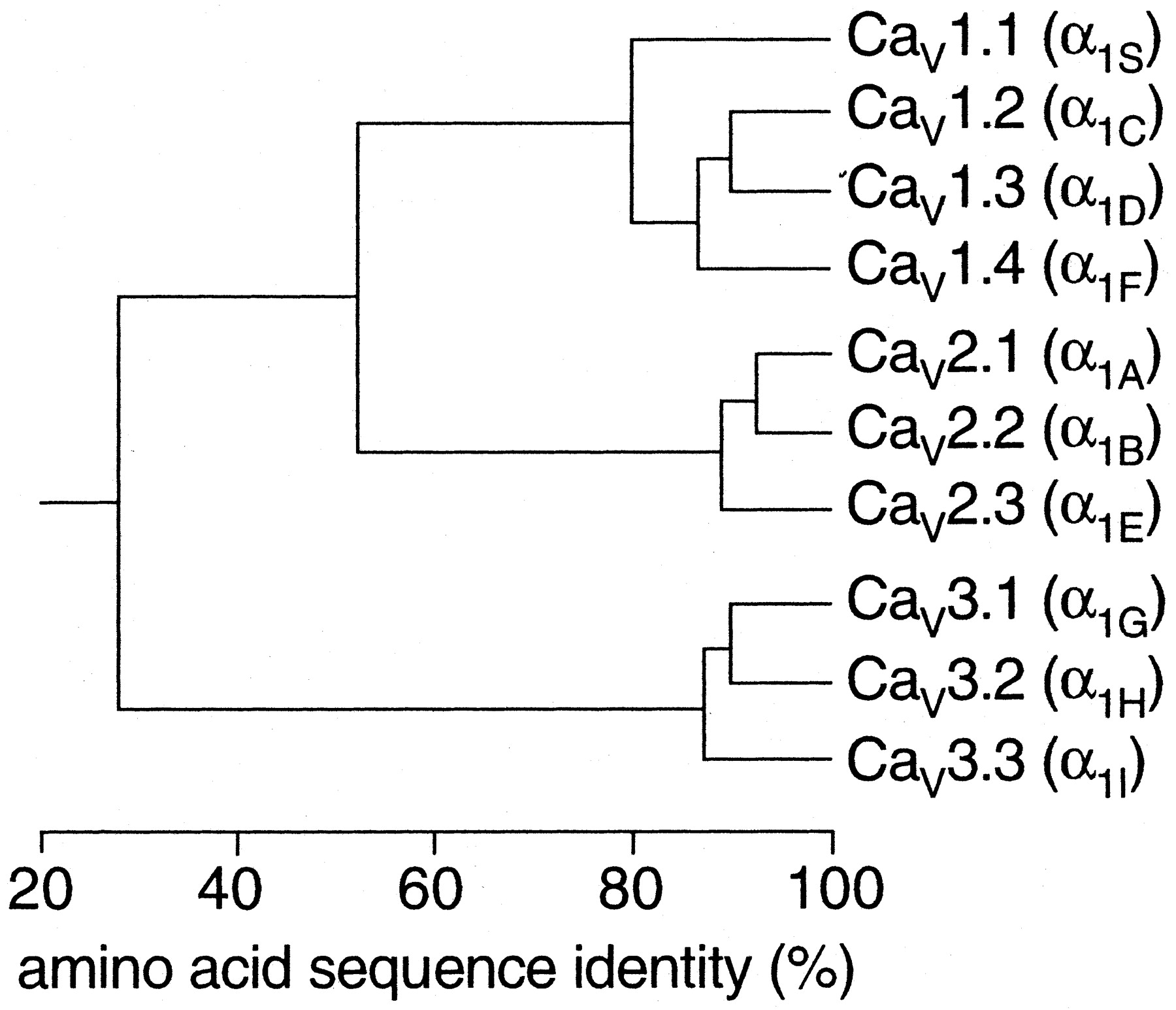
The complete amino acid sequences of these {alpha}1 subunits are more than 70% identical within a family but less than 40% identical among families. These family relationships are illustrated for the more conserved transmembrane and pore domains in Fig. 2. Division of calcium channels into these three families is phylogenetically ancient, as representatives of each are found in the Caenorhabditis elegans genome. Consequently, the genes for the different {alpha}1 subunits have become widely dispersed in the genome and even the most closely related members of the family are not clustered on single chromosomes.


View larger version (28K):
[in this window]
[in a new window]
 
FIG. 2. Sequence similarity of voltage-gated calcium channel {alpha}1 subunits. Phylogenetic representation of the primary sequences of the calcium channels. Only the membrane-spanning segments and the pore loops (~350 amino acids) are compared. First, all sequence pairs were compared, which clearly defines three families with intra-family sequence identities above 80% (CaV1, CaV2, CaV3). Then, a consensus sequence was defined for each family and these three sequences were compared with one another, with inter-family sequence identities of ~52% (CaV1 versus CaV2) and 28% (CaV3 versus and CaV1 or CaV2).


    Calcium Channel Molecular Pharmacology
 Top
 Previous
 Next
 
The pharmacology of the three families of calcium channel is quite distinct. The CaV1 channels are the molecular targets of the organic calcium channel blockers used widely in the therapy of cardiovascular diseases. These drugs are thought to act at three separate, but allosterically coupled, receptor sites (Table 1). Phenylalkylamines are intracellular pore blockers, which are thought to enter the pore from the cytoplasmic side of the channel and block it. Their receptor site is formed by amino acid residues in the S6 segments in domains III and IV, in close analogy to the local anesthetic receptor site on sodium channels (Hockerman et al., 1997Go; Hofmann et al., 1999Go; Striessnig, 1999Go). Dihydropyridines can be channel activators or inhibitors and therefore are thought to act allosterically to shift the channel toward the open or closed state, rather than by occluding the pore. Their receptor site includes amino acid residues in the S6 segments of domains III and IV and the S5 segment of domain III. The dihydropyridine receptor site is closely apposed to the phenylalkylamine receptor site and shares some common amino acid residues. Diltiazem and related benzothiazepines are thought to bind to a third receptor site, but the amino acid residues that are required for their binding overlap extensively with those required for phenylalkylamine binding.

The CaV2 family of calcium channels is relatively insensitive to dihydropyridine calcium channel blockers, but these calcium channels are specifically blocked with high affinity by peptide toxins from spiders and marine snails (Miljanich and Ramachandran, 1995Go). The CaV2.1 channels are blocked specifically by {omega}-agatoxin IVA from funnel web spider venom. The CaV2.2 channels are blocked specifically by {omega}-conotoxin GVIA and related cone snail toxins. The CaV2.3 channels are blocked specifically by the synthetic peptide toxin SNX-482 derived from tarantula venom. These peptide toxins are potent blockers of synaptic transmission because of their specific effects on the CaV2 family of calcium channels.

The CaV3 family of calcium channels is insensitive to both the dihydropyridines that block CaV1 channels and the spider and cone snail toxins that block the CaV2 channels, and there are no widely useful pharmacological agents that block T-type calcium currrents (Heady et al., 2001Go). The organic calcium channel blockers mibefradil is somewhat specific for T-type versus L-type calcium currents (3- to 5-fold). The peptide kurtoxin inhibits the activation gating of CaV3.1 and CaV3.2 channels. Development of more specific and high affinity blockers of the CaV3 family of calcium channels would be useful for therapy and for more detailed analysis of the physiological roles of these channels.

This section of the compendium summarizes the major molecular, physiological, and pharmacological properties for each of the ten calcium channels that have been functionally expressed. Quantitative data are included for voltage dependence of activation and inactivation, single-channel conductance, and binding of drugs and neurotoxins, focusing on those agents that are widely used and are diagnostic of channel identity and function.


    Footnotes
 
Address correspondence to: Dr. William A. Catterall, Department of Pharmacology, University of Washington School of Medicine, Box 357280, Seattle, WA 98195-7280. E-mail: wcatt{at}u.washington.edu

DOI: 10.1124/pr.55.4.8.

1 This work was previously published in Catterall WA, Chandy KG, and Gutman GA, eds. (2002) The IUPHAR Compendium of Voltage-Gated Ion Channels, International Union of Pharmacology Media, Leeds, UK. Back


    References
 Top
 

Birnbaumer L, Campbell KP, Catterall WA, Harpold MM, Hofmann F, Horne WA, Mori Y, Schwartz A, Snutch TP, Tanabe T, et al. (1994) The naming of voltage-gated calcium channels. Neuron 13: 505-506.[CrossRef][Medline]

Catterall WA (1995) Structure and function of voltage-gated ion channels. Annu Rev Biochem 65: 493-531.[CrossRef]

Catterall WA (2000) Structure and regulation of voltage-gated Ca2+ channels. Annu Rev Cell Dev Biol 16: 521-555.[CrossRef][Medline]

Chandy KG and Gutman GA (1993) Nomenclature for mammalian potassium channel genes. Trends Pharmacol Sci 14: 434.[CrossRef][Medline]

Ertel EA, Campbell KP, Harpold MM, Hofmann F, Mori Y, Perez-Reyes E, Schwartz A, Snutch TP, Tanabe T, Birnbaumer L, et al. (2000) Nomenclature of voltage-gated calcium channels. Neuron 25: 533-535.[CrossRef][Medline]

Glossmann H and Striessnig J (1990) Molecular properties of calcium channels. Rev Physiol Biochem Pharmacol 114: 1-105.[Medline]

Heady TN, Gomora JC, Macdonald TL, and Perez-Reyes E (2001) Molecular pharmacology of T-type Ca2+ channels. Jpn J Pharmacol 85: 339-350.[CrossRef][Medline]

Hockerman GH, Peterson BZ, Johnson BD, and Catterall WA (1997) Molecular determinants of drug binding and action on L-type calcium channels. Annu Rev Pharmacol Toxicol 37: 361-396.[CrossRef][Medline]

Hofmann F, Biel M, and Flockerzi V (1994) Molecular basis for Ca2+ channel diversity. Annu Rev Neurosci 17: 399-418.[CrossRef][Medline]

Hofmann F, Lacinová L, and Klugbauer N (1999) Voltage-dependent calcium channels: from structure to function. Rev Physiol Biochem Pharmacol 139: 33-87.[Medline]

Miljanich GP and Ramachandran J (1995) Antagonists of neuronal calcium channels: structure, function, and therapeutic implications. Annu Rev Pharmacol Toxicol 35: 707-734.[CrossRef][Medline]

Striessnig J (1999) Pharmacology, structure and function of cardiac L-type calcium channels. Cell Physiol Biochem 9: 242-269.[Medline]

Tsien RW, Lipscombe D, Madison D, Bley K, and Fox A (1995) Reflections on calcium channel diversity, 1988–1994. Trends Neurosci 18: 52-54.[CrossRef][Medline]


This article has been cited by other articles:


Home page
Physiol. GenomicsHome page
S. Ellefsen, G. K. Sandvik, H. K. Larsen, K.-O. Stenslokken, D. A. S. Hov, T. A. Kristensen, and G. E. Nilsson
Expression of genes involved in excitatory neurotransmission in anoxic crucian carp (Carassius carassius) brain
Physiol Genomics, September 17, 2008; 35(1): 5 - 17.
[Abstract] [Full Text] [PDF]


Home page
Physiol. Rev.Home page
M. E. Mangoni and J. Nargeot
Genesis and Regulation of the Heart Automaticity
Physiol Rev, July 1, 2008; 88(3): 919 - 982.
[Abstract] [Full Text] [PDF]


Home page
J. Dent. Res.Home page
G. Chung, J.N. Rhee, S.J. Jung, J.S. Kim, and S.B. Oh
Modulation of CaV2.3 Calcium Channel Currents by Eugenol
J. Dent. Res., February 1, 2008; 87(2): 137 - 141.
[Abstract] [Full Text] [PDF]


Home page
J. Physiol.Home page
B. K. Rycroft, K. S. Vikman, and M. J. Christie
Inflammation reduces the contribution of N-type calcium channels to primary afferent synaptic transmission onto NK1 receptor-positive lamina I neurons in the rat dorsal horn
J. Physiol., May 1, 2007; 580(3): 883 - 894.
[Abstract] [Full Text] [PDF]


Home page
Am. J. Physiol. Heart Circ. Physiol.Home page
G. Baroudi, Y. Qu, O. Ramadan, M. Chahine, and M. Boutjdir
Protein kinase C activation inhibits Cav1.3 calcium channel at NH2-terminal serine 81 phosphorylation site.
Am J Physiol Heart Circ Physiol, October 1, 2006; 291(4): H1614 - H1622.
[Abstract] [Full Text] [PDF]


Home page
Am. J. Physiol. Cell Physiol.Home page
M. C. Chen, S. V. Wu, J. R. Reeve Jr., and E. Rozengurt
Bitter stimuli induce Ca2+ signaling and CCK release in enteroendocrine STC-1 cells: role of L-type voltage-sensitive Ca2+ channels
Am J Physiol Cell Physiol, October 1, 2006; 291(4): C726 - C739.
[Abstract] [Full Text] [PDF]


Home page
Nucleic Acids ResHome page
R. Johnson, R. J. Gamblin, L. Ooi, A. W. Bruce, I. J. Donaldson, D. R. Westhead, I. C. Wood, R. M. Jackson, and N. J. Buckley
Identification of the REST regulon reveals extensive transposable element-mediated binding site duplication
Nucleic Acids Res., September 1, 2006; 34(14): 3862 - 3877.
[Abstract] [Full Text] [PDF]


Home page
J. Biol. Chem.Home page
T.-S. Lee, R. Karl, S. Moosmang, P. Lenhardt, N. Klugbauer, F. Hofmann, T. Kleppisch, and A. Welling
Calmodulin Kinase II Is Involved in Voltage-dependent Facilitation of the L-type Cav1.2 Calcium Channel: IDENTIFICATION OF THE PHOSPHORYLATION SITES
J. Biol. Chem., September 1, 2006; 281(35): 25560 - 25567.
[Abstract] [Full Text] [PDF]


Home page
Am. J. Physiol. Gastrointest. Liver Physiol.Home page
E. Rozengurt
Taste Receptors in the Gastrointestinal Tract. I. Bitter taste receptors and {alpha}-gustducin in the mammalian gut
Am J Physiol Gastrointest Liver Physiol, August 1, 2006; 291(2): G171 - G177.
[Abstract] [Full Text] [PDF]


Home page
J AndrolHome page
R. A. Anderson, K. A. Feathergill, D. P. Waller, and L. J. D. Zaneveld
SAMMA Induces Premature Human Acrosomal Loss by Ca2+ Signaling Dysregulation
J Androl, July 1, 2006; 27(4): 568 - 577.
[Abstract] [Full Text] [PDF]


Home page
J. Pharmacol. Exp. Ther.Home page
Y.-J. Won, K. Whang, I. D. Kong, K.-S. Park, J.-W. Lee, and S.-W. Jeong
Expression Profiles of High Voltage-Activated Calcium Channels in Sympathetic and Parasympathetic Pelvic Ganglion Neurons Innervating the Urogenital System
J. Pharmacol. Exp. Ther., June 1, 2006; 317(3): 1064 - 1071.
[Abstract] [Full Text] [PDF]


Home page
EndocrinologyHome page
J. Hall, R. D. Jones, T. H. Jones, K. S. Channer, and C. Peers
Selective Inhibition of L-Type Ca2+ Channels in A7r5 Cells by Physiological Levels of Testosterone
Endocrinology, June 1, 2006; 147(6): 2675 - 2680.
[Abstract] [Full Text] [PDF]


Home page
Hum Reprod UpdateHome page
C. Jimenez-Gonzalez, F. Michelangeli, C.V. Harper, C.L.R. Barratt, and S.J. Publicover
Calcium signalling in human spermatozoa: a specialized 'toolkit' of channels, transporters and stores
Hum. Reprod. Update, May 1, 2006; 12(3): 253 - 267.
[Abstract] [Full Text] [PDF]


Home page
J. Biol. Chem.Home page
A. Hohaus, S. Beyl, M. Kudrnac, S. Berjukow, E. N. Timin, R. Marksteiner, M. A. Maw, and S. Hering
Structural Determinants of L-type Channel Activation in Segment IIS6 Revealed by a Retinal Disorder
J. Biol. Chem., November 18, 2005; 280(46): 38471 - 38477.
[Abstract] [Full Text] [PDF]


Home page
J. Clin. Endocrinol. Metab.Home page
B. Liu, S. J. Hill, and R. N. Khan
Oxytocin Inhibits T-Type Calcium Current of Human Decidual Stromal Cells
J. Clin. Endocrinol. Metab., July 1, 2005; 90(7): 4191 - 4197.
[Abstract] [Full Text] [PDF]


Home page
Br J AnaesthHome page
I. L. White, N. P. Franks, and R. Dickinson
Effects of isoflurane and xenon on Ba2+-currents mediated by N-type calcium channels
Br. J. Anaesth., June 1, 2005; 94(6): 784 - 790.
[Abstract] [Full Text] [PDF]


Home page
ReproductionHome page
R. Felix
Molecular physiology and pathology of Ca2+-conducting channels in the plasma membrane of mammalian sperm
Reproduction, March 1, 2005; 129(3): 251 - 262.
[Abstract] [Full Text] [PDF]


Home page
J. Neurosci.Home page
S. P. Brown, P. K. Safo, and W. G. Regehr
Endocannabinoids Inhibit Transmission at Granule Cell to Purkinje Cell Synapses by Modulating Three Types of Presynaptic Calcium Channels
J. Neurosci., June 16, 2004; 24(24): 5623 - 5631.
[Abstract] [Full Text] [PDF]


Home page
J. Biol. Chem.Home page
L. Stokes, J. Gordon, and G. Grafton
Non-voltage-gated L-type Ca2+ Channels in Human T Cells: PHARMACOLOGY AND MOLECULAR CHARACTERIZATION OF THE MAJOR {alpha} PORE-FORMING AND AUXILIARY {beta}-SUBUNITS
J. Biol. Chem., May 7, 2004; 279(19): 19566 - 19573.
[Abstract] [Full Text] [PDF]


This Article
Right arrow Abstract Freely available
Right arrow Full Text (PDF)
Right arrow Submit a response
Right arrow Alert me when this article is cited
Right arrow Alert me when eLetters are posted
Right arrow Alert me if a correction is posted
Services
Right arrow Similar articles in this journal
Right arrow Similar articles in PubMed
Right arrow Alert me to new issues of the journal
Right arrow Download to citation manager
Citing Articles
Right arrow Citing Articles via HighWire
Right arrow Citing Articles via Google Scholar
Google Scholar
Right arrow Articles by Catterall, W. A.
Right arrow Articles by Perez-Reyes, E.
Right arrow Search for Related Content
PubMed
Right arrow PubMed Citation
Right arrow Articles by Catterall, W. A.
Right arrow Articles by Perez-Reyes, E.


HOME HELP FEEDBACK SUBSCRIPTIONS ARCHIVE SEARCH TABLE OF CONTENTS
All ASPET Journals Molecular Pharmacology Pharmacological Reviews
 Molecular Interventions Drug Metabolism and Disposition
Copyright © 2003 by the American Society for Pharmacology and Experimental Therapeutics