Advertisement

Main menu

  • Home
  • Articles
    • Current Issue
    • Latest Articles
    • Archive
  • Information
    • Instructions to Authors
    • Submit a Manuscript
    • FAQs
    • For Subscribers
    • Terms & Conditions of Use
    • Permissions
  • Editorial Board
  • Alerts
    • Alerts
    • RSS Feeds
  • Virtual Issues
  • Feedback
  • Other Publications
    • Drug Metabolism and Disposition
    • Journal of Pharmacology and Experimental Therapeutics
    • Molecular Pharmacology
    • Pharmacological Reviews
    • Pharmacology Research & Perspectives
    • ASPET

User menu

  • My alerts
  • Log in
  • My Cart

Search

  • Advanced search
Pharmacological Reviews
  • Other Publications
    • Drug Metabolism and Disposition
    • Journal of Pharmacology and Experimental Therapeutics
    • Molecular Pharmacology
    • Pharmacological Reviews
    • Pharmacology Research & Perspectives
    • ASPET
  • My alerts
  • Log in
  • My Cart
Pharmacological Reviews

Advanced Search

  • Home
  • Articles
    • Current Issue
    • Latest Articles
    • Archive
  • Information
    • Instructions to Authors
    • Submit a Manuscript
    • FAQs
    • For Subscribers
    • Terms & Conditions of Use
    • Permissions
  • Editorial Board
  • Alerts
    • Alerts
    • RSS Feeds
  • Virtual Issues
  • Feedback
  • Visit Pharm Rev on Facebook
  • Follow Pharm Rev on Twitter
  • Follow ASPET on LinkedIn
OtherIUPHAR Nomenclature Report
Open Access

International Union of Basic and Clinical Pharmacology. CVII. Structure and Pharmacology of the Apelin Receptor with a Recommendation that Elabela/Toddler Is a Second Endogenous Peptide Ligand

Cai Read, Duuamene Nyimanu, Thomas L. Williams, David J. Huggins, Petra Sulentic, Robyn G. C. Macrae, Peiran Yang, Robert C. Glen, Janet J. Maguire and Anthony P. Davenport
Eliot H. Ohlstein, ASSOCIATE EDITOR
Pharmacological Reviews October 2019, 71 (4) 467-502; DOI: https://doi.org/10.1124/pr.119.017533
Cai Read
Experimental Medicine and Immunotherapeutics, University of Cambridge, Centre for Clinical Investigation, Addenbrooke’s Hospital, Cambridge, United Kingdom (C.R., D.N., T.L.W., D.J.H., P.S., R.G.C.M., P.Y., J.J.M., A.P.D.); The Centre for Molecular Informatics, Department of Chemistry, University of Cambridge, Cambridge, United Kingdom (D.J.H., R.C.G.); and Computational and Systems Medicine, Department of Surgery and Cancer, Faculty of Medicine, Imperial College London, London, United Kingdom (R.C.G.)
  • Find this author on Google Scholar
  • Find this author on PubMed
  • Search for this author on this site
Duuamene Nyimanu
Experimental Medicine and Immunotherapeutics, University of Cambridge, Centre for Clinical Investigation, Addenbrooke’s Hospital, Cambridge, United Kingdom (C.R., D.N., T.L.W., D.J.H., P.S., R.G.C.M., P.Y., J.J.M., A.P.D.); The Centre for Molecular Informatics, Department of Chemistry, University of Cambridge, Cambridge, United Kingdom (D.J.H., R.C.G.); and Computational and Systems Medicine, Department of Surgery and Cancer, Faculty of Medicine, Imperial College London, London, United Kingdom (R.C.G.)
  • Find this author on Google Scholar
  • Find this author on PubMed
  • Search for this author on this site
Thomas L. Williams
Experimental Medicine and Immunotherapeutics, University of Cambridge, Centre for Clinical Investigation, Addenbrooke’s Hospital, Cambridge, United Kingdom (C.R., D.N., T.L.W., D.J.H., P.S., R.G.C.M., P.Y., J.J.M., A.P.D.); The Centre for Molecular Informatics, Department of Chemistry, University of Cambridge, Cambridge, United Kingdom (D.J.H., R.C.G.); and Computational and Systems Medicine, Department of Surgery and Cancer, Faculty of Medicine, Imperial College London, London, United Kingdom (R.C.G.)
  • Find this author on Google Scholar
  • Find this author on PubMed
  • Search for this author on this site
David J. Huggins
Experimental Medicine and Immunotherapeutics, University of Cambridge, Centre for Clinical Investigation, Addenbrooke’s Hospital, Cambridge, United Kingdom (C.R., D.N., T.L.W., D.J.H., P.S., R.G.C.M., P.Y., J.J.M., A.P.D.); The Centre for Molecular Informatics, Department of Chemistry, University of Cambridge, Cambridge, United Kingdom (D.J.H., R.C.G.); and Computational and Systems Medicine, Department of Surgery and Cancer, Faculty of Medicine, Imperial College London, London, United Kingdom (R.C.G.)
  • Find this author on Google Scholar
  • Find this author on PubMed
  • Search for this author on this site
Petra Sulentic
Experimental Medicine and Immunotherapeutics, University of Cambridge, Centre for Clinical Investigation, Addenbrooke’s Hospital, Cambridge, United Kingdom (C.R., D.N., T.L.W., D.J.H., P.S., R.G.C.M., P.Y., J.J.M., A.P.D.); The Centre for Molecular Informatics, Department of Chemistry, University of Cambridge, Cambridge, United Kingdom (D.J.H., R.C.G.); and Computational and Systems Medicine, Department of Surgery and Cancer, Faculty of Medicine, Imperial College London, London, United Kingdom (R.C.G.)
  • Find this author on Google Scholar
  • Find this author on PubMed
  • Search for this author on this site
Robyn G. C. Macrae
Experimental Medicine and Immunotherapeutics, University of Cambridge, Centre for Clinical Investigation, Addenbrooke’s Hospital, Cambridge, United Kingdom (C.R., D.N., T.L.W., D.J.H., P.S., R.G.C.M., P.Y., J.J.M., A.P.D.); The Centre for Molecular Informatics, Department of Chemistry, University of Cambridge, Cambridge, United Kingdom (D.J.H., R.C.G.); and Computational and Systems Medicine, Department of Surgery and Cancer, Faculty of Medicine, Imperial College London, London, United Kingdom (R.C.G.)
  • Find this author on Google Scholar
  • Find this author on PubMed
  • Search for this author on this site
Peiran Yang
Experimental Medicine and Immunotherapeutics, University of Cambridge, Centre for Clinical Investigation, Addenbrooke’s Hospital, Cambridge, United Kingdom (C.R., D.N., T.L.W., D.J.H., P.S., R.G.C.M., P.Y., J.J.M., A.P.D.); The Centre for Molecular Informatics, Department of Chemistry, University of Cambridge, Cambridge, United Kingdom (D.J.H., R.C.G.); and Computational and Systems Medicine, Department of Surgery and Cancer, Faculty of Medicine, Imperial College London, London, United Kingdom (R.C.G.)
  • Find this author on Google Scholar
  • Find this author on PubMed
  • Search for this author on this site
Robert C. Glen
Experimental Medicine and Immunotherapeutics, University of Cambridge, Centre for Clinical Investigation, Addenbrooke’s Hospital, Cambridge, United Kingdom (C.R., D.N., T.L.W., D.J.H., P.S., R.G.C.M., P.Y., J.J.M., A.P.D.); The Centre for Molecular Informatics, Department of Chemistry, University of Cambridge, Cambridge, United Kingdom (D.J.H., R.C.G.); and Computational and Systems Medicine, Department of Surgery and Cancer, Faculty of Medicine, Imperial College London, London, United Kingdom (R.C.G.)
  • Find this author on Google Scholar
  • Find this author on PubMed
  • Search for this author on this site
Janet J. Maguire
Experimental Medicine and Immunotherapeutics, University of Cambridge, Centre for Clinical Investigation, Addenbrooke’s Hospital, Cambridge, United Kingdom (C.R., D.N., T.L.W., D.J.H., P.S., R.G.C.M., P.Y., J.J.M., A.P.D.); The Centre for Molecular Informatics, Department of Chemistry, University of Cambridge, Cambridge, United Kingdom (D.J.H., R.C.G.); and Computational and Systems Medicine, Department of Surgery and Cancer, Faculty of Medicine, Imperial College London, London, United Kingdom (R.C.G.)
  • Find this author on Google Scholar
  • Find this author on PubMed
  • Search for this author on this site
Anthony P. Davenport
Experimental Medicine and Immunotherapeutics, University of Cambridge, Centre for Clinical Investigation, Addenbrooke’s Hospital, Cambridge, United Kingdom (C.R., D.N., T.L.W., D.J.H., P.S., R.G.C.M., P.Y., J.J.M., A.P.D.); The Centre for Molecular Informatics, Department of Chemistry, University of Cambridge, Cambridge, United Kingdom (D.J.H., R.C.G.); and Computational and Systems Medicine, Department of Surgery and Cancer, Faculty of Medicine, Imperial College London, London, United Kingdom (R.C.G.)
  • Find this author on Google Scholar
  • Find this author on PubMed
  • Search for this author on this site
Eliot H. Ohlstein
Roles: ASSOCIATE EDITOR
  • Find this author on Google Scholar
  • Find this author on PubMed
  • Search for this author on this site
  • Article
  • Figures & Data
  • Info & Metrics
  • eLetters
  • PDF
Loading

Article Figures & Data

Figures

  • Tables
  • Fig. 1.
    • Download figure
    • Open in new tab
    • Download powerpoint
    Fig. 1.

    Predicted disulfide bridges are between Cys19–Cys281 and Cys102–Cys181 (yellow); glycosylation sites (blue) are in the N-terminal tail (Asn15) and extracellular loop 2 (ECL2; Asn175); palmitoylation site (green) Cys325 and Cys326 and phosphorylation site (purple) Ser348 have been confirmed experimentally, of which Ser348 is crucial for apelin receptor interactions with GRK2/5, β-arrestin, and its internalization (Chen et al., 2014). Figure constructed from G protein-coupled receptor database (Pándy-Szekeres et al., 2018).
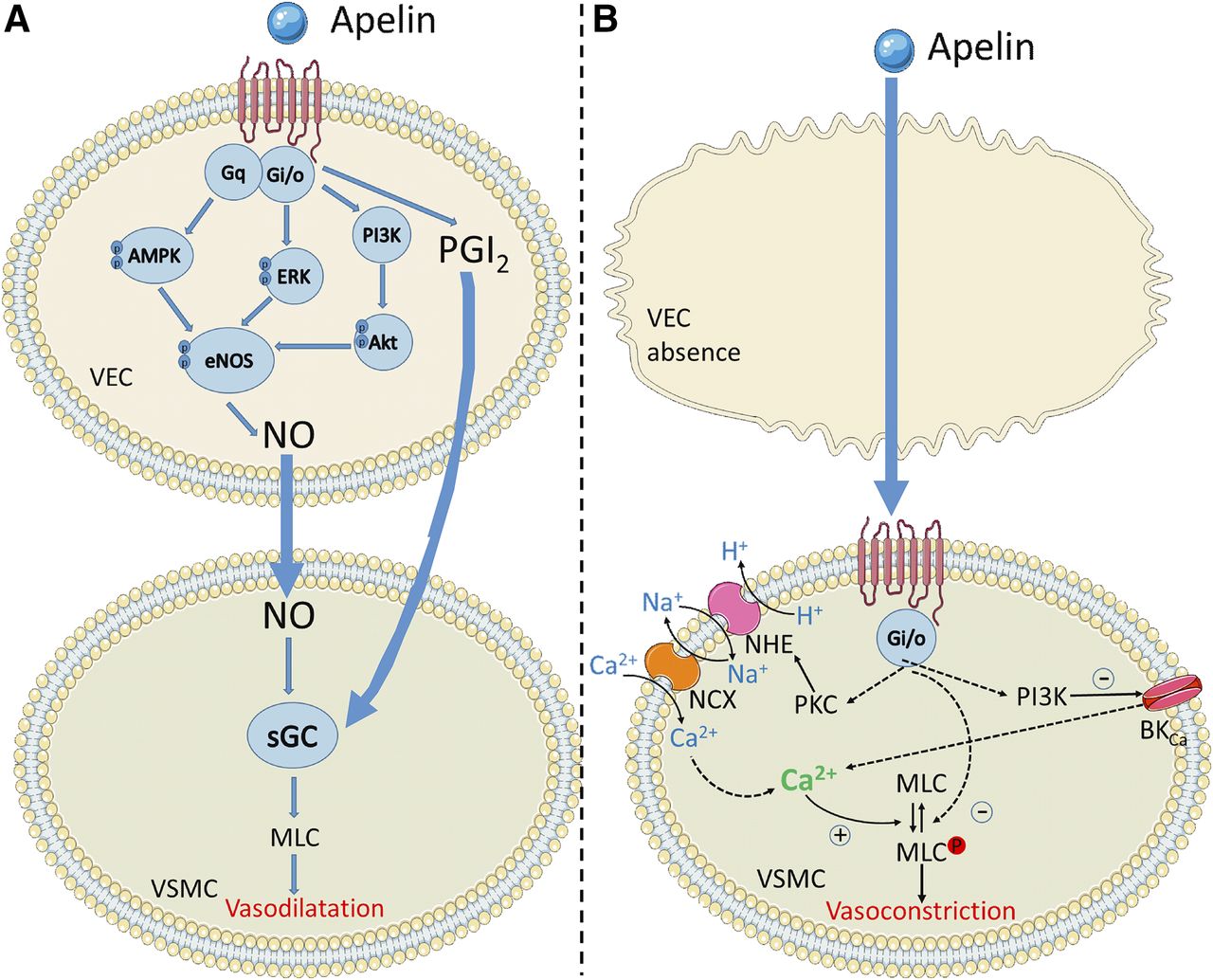
  • Fig. 2.
    • Download figure
    • Open in new tab
    • Download powerpoint
    Fig. 2.

    The key signaling pathways suspected to be activated in vascular endothelial cells (VEC) and smooth muscle cells (VSMC) by the apelin receptor. Apelin binding can promote Gαi, Gαq, and β-arrestin recruitment to the receptor. In the presence of the endothelium, both Gαi and Gαq promote relaxation of smooth muscle cells through nitric oxide and prostacyclin release. In the absence of the endothelium, apelin binds directly to the receptor on the smooth muscle cells and leads to constriction through undetermined intermediate steps but most likely involving PKC, phosphoinositide 3-kinase (PI3K) and myosin light chain phosphorylation. Figure constructed using Servier Medical Art.
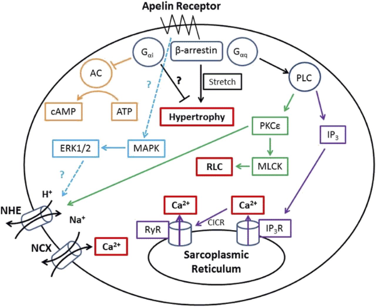
  • Fig. 3.
    • Download figure
    • Open in new tab
    • Download powerpoint
    Fig. 3.

    The key signaling pathways suspected to be activated in cardiomyocytes by the apelin receptor. Apelin binding can promote Gαi, Gαq, and β-arrestin recruitment to the receptor, these pathways are thought to ultimately lead to cardiac inotropy without hypertrophy. However, in the absence of apelin, β-arrestin recruitment may lead to stretch-mediated hypertrophy.

  • Fig. 4.
    • Download figure
    • Open in new tab
    • Download powerpoint
    Fig. 4.

    An overlay of ELA-11 (green) and apelin-13 (blue) docked in the apelin receptor binding pocket. The peptide sequences are shown alongside with the same color scheme. The red amino acids show where identical residues line up. Overlay from Yang et al., (2017b) under CC-BY license.

  • Fig. 5.
    • Download figure
    • Open in new tab
    • Download powerpoint
    Fig. 5.

    The amino acid sequences of cleaved apelin fragments. [Pyr1]apelin-13 is the predominant form in the cardiovascular system and is shown in red with the pyroglutamate residue in pink. The smallest active fragment is highlighted, as well as the RPRL motif which has been thought critical to binding.

  • Fig. 6.
    • Download figure
    • Open in new tab
    • Download powerpoint
    Fig. 6.

    The amino acid sequences of the predicted cleaved ELA fragments compared with [Pyr1]apelin-13, the predominant apelin isoform in the cardiovascular system. There is little sequence homology between ELA and apelin fragments; however, there are some similarities in the positioning of charged residues. Disulfide bridges are yellow lines, hydrophobic amino acids are shown in green, uncharged polar amino acids in pink, basic amino acids in blue and pyroglutamate in red. From Yang et al. (2017b) under CC-BY license.
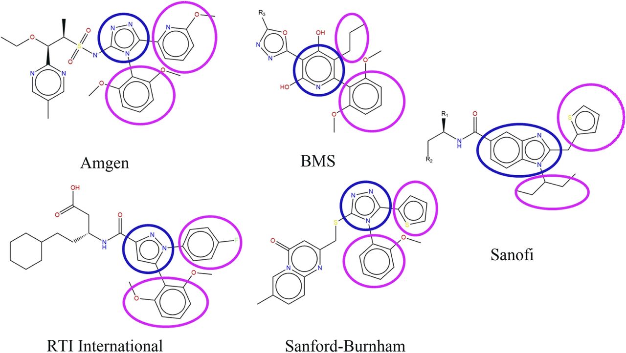
  • Fig. 7.
    • Download figure
    • Open in new tab
    • Download powerpoint
    Fig. 7.

    The scaffold structures of five reported series of small molecule apelin agonists from Amgen (Chen et al., 2017), Bristol-Myers Squibb (Myers et al., 2017), RTI International (Narayanan et al., 2016), Sanford-Burnham (Pinkerton and Smith, 2015), and Sanofi (Hachtel et al., 2014). CMF-019 is derived from the Sanofi series. All of these molecules possess a broadly similar structure, consisting of two hydrophobic groups (circled in pink) extending from a heterocyclic core group (in blue).
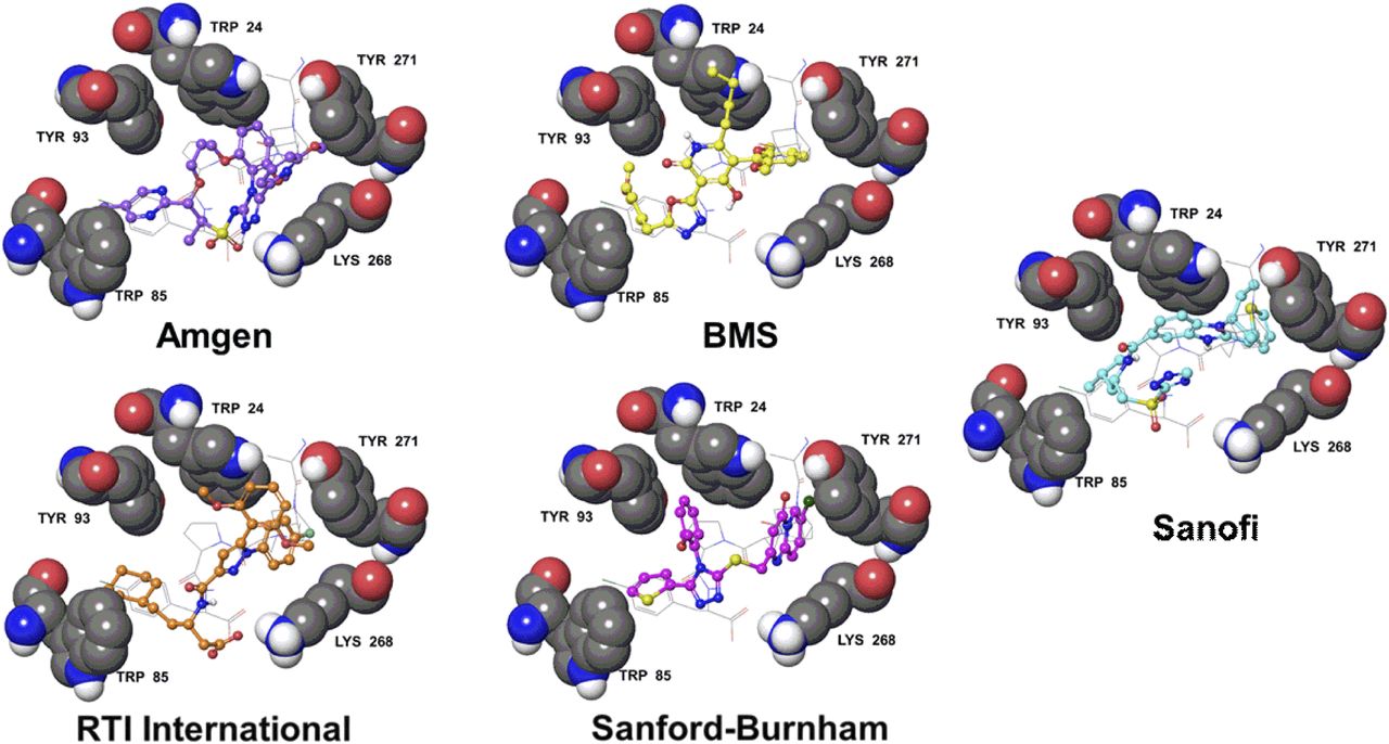
  • Fig. 8.
    • Download figure
    • Open in new tab
    • Download powerpoint
    Fig. 8.

    The apelin receptor structure from PDBID 5VBL (Ma et al., 2017) with docked poses of five series of apelin agonists. The apelin receptor residues, W24, W85, Y93, K268, and Y271 are labeled and displayed as gray space filling. The structure of the four C-terminal residues of the apelin analog from PDBID 5VBL are displayed as gray sticks. The receptor and peptide are overlaid with docked poses of an Amgen (violet balls and sticks), a Bristol-Myers Squibb (yellow balls and sticks), an RTI International (orange balls and sticks), a Sanford-Burnham (magenta balls and sticks), and a Sanofi (cyan balls and sticks) small molecule apelin agonist. Only polar hydrogens are shown. Structures were derived from the following patents: Amgen (Chen et al., 2017), Bristol-Myers Squibb (Myers et al., 2017), RTI International (Narayanan et al., 2016), Sanford-Burnham (Pinkerton and Smith, 2015), and Sanofi (Hachtel et al., 2014). Pinkerton and Smith (2015) confirmed the Sanford-Burnham apelin compounds were selective vs. the angiotensin II receptor (AT1), the most closely related GPCR, with no significant off target binding.

Tables

  • Figures
    • View popup
    TABLE 1

    Contacts between apelin-13 and the apelin receptor, inferred from the crystal structure PDBID 5VBL

    Apelin-13 ContactReceptor ContactsReferences
    Q1 SidechainD92aGerbier et al., 2015
    Q1 BackboneN177
    P3 SidechainT22
    D23aZhou et al., 2003a
    R4 BackboneD23aZhou et al., 2003a
    L5 SidechainE20aZhou et al., 2003a
    T22
    S6 SidechainE174aChapman et al., 2014; Gerbier et al., 2015
    S6 BackboneY21
    H7 SidechainE174aChapman et al., 2014; Gerbier et al., 2015
    K8 SidechainY21Gerbier et al., 2015
    D284aMa et al., 2017
    S275
    G9 backboneY271
    P10 SidechainE198a
    L173Chapman et al., 2014
    M11 SidechainW24
    Y271aMa et al., 2017
    F291
    K268bKumar et al., 2016
    P12 SidechainW24
    Y93
    P12 BackboneR168aMa et al., 2017
    F13 SidechainW85a
    Y88
    T89
    Y93
    Y299
    F13 CarboxylateK268bKumar et al., 2016
    Y264
    • ↵a Receptor residues implicated in apelin binding by mutagenesis.

    • ↵b Receptor residues affecting bias and internalization by mutagenesis.

    • View popup
    TABLE 2

    Expression of apelin receptor (APLNR), apelin, and ELA in mouse, rat, and human tissues

    Compiled from Edinger et al. (1998), Hosoya et al. (2000), Lee et al. (2000), O’Carroll et al. (2000), Medhurst et al. (2003), Kleinz and Davenport (2005), Regard et al. (2008), Pope et al. (2012), Deng et al. (2015), Wang et al. (2015b).

    APLNRApelinELA
    HumanRatMouseHumanRatMouseHumanRatMouse
    Brain++++++++++++
    -paraventricular nucleus+++
    -supraoptic nucleus++++
    Anterior pituitary+a++++++++a+a
    Intermediate pituitary−/+−/++/+++
    Posterior pituitary−/+−/+++/++
    Thymus+−+−
    Spinal cord++++++++++
    Cerebellum++−++−
    Hippocampus++−++
    Thalamus++++
    Heart+++++++++++−/++
    Adrenal gland+++
    Lungs++++++++++++++
    Stomach+++−+
    Liver+−+−−−
    Small intestine++++−+
    Large intestine+++++
    Pancreas+−++
    Kidney+++++++++++
    Testis+++++++
    Prostate++++
    Ovary++++++
    Uterus++++−+−
    Placenta++++++
    Mammary gland+++
    Skeletal muscle+++++−++
    Adipose tissue++++++b++
    Cartilage++++
    Spleen+++−+−+
    Skin++
    Bladder++
    Gall bladder+
    • +++, high expression; ++, moderate expression; low expression; −, absence mRNA expression.

    • ↵a Expression in whole pituitary gland.

    • ↵b Expression reported in both white and brown adipose tissue.

    • View popup
    TABLE 3

    Some of the key agonists at the apelin receptor, their binding affinities, and whether they demonstrate bias compared with [Pyr1]apelin-13 (the predominant apelin isoform in the cardiovascular system (Maguire el al., 2009; Zhen et al., 2013) Endogenous agonists are denoted by “(E).”

    LigandActionBinding AffinityUnitsBiasReferences
    [Pyr1]Apelin-13 (E)Full Agonist7.0–8.8pIC50—Kawamata et al., 2001
    Medhurst et al., 2003
    Apelin-13 (E)Full Agonist8.8–9.5pIC50—Fan et al., 2003
    Hosoya et al., 2000
    Medhurst et al., 2003
    Apelin-17 (E)Full Agonist7.9–9.0pIC50β-arrestinEl Messari et al., 2004
    Medhurst et al., 2003
    Apelin-36 (E)Full Agonist8.2–8.6pIC50—Fan et al., 2003
    Hosoya et al., 2000
    Kawamata et al., 2001
    Medhurst et al., 2003
    Elabela/Toddler-11 (E)Full Agonist7.2pIC50Yang et al., 2016
    Elabela/Toddler-21 (E)Full Agonist8.7pIC50β-arrestinYang et al., 2016
    Elabela/Toddler-32 (E)Full Agonist8.7pIC50β-arrestinYang et al., 2016
    MM07Full Agonist9.5pEC50G proteinBrame et al., 2015
    CMF-019Full Agonist8.6pIC50G proteinRead et al., 2016
    ML233Full Agonist———Khan et al., 2011
    E339-3D6Full Agonist6.4pKi—Iturrioz et al., 2010a
    • View popup
    TABLE 4

    Some of the key antagonists at the apelin receptor and their binding affinities

    MM54, an antagonist at the β-arrestin and internalization pathway, consists of a cyclized peptide based around the RPRL motif. MM54 has been tested for selectivity (Bowes et al., 2012) against over 50 GPCRs (including the most closely related angiotensin II receptor AT1) and ion channels. ALX40-4C and protamine both consist of a series of positively charged amino acids and display low binding affinities and likely low selectivity for the apelin receptor.

    LigandActionBinding AffinityUnitsReferences
    MM54Antagonist8.2pKiMacaluso et al., 2011
    ALX40-4CAntagonist5.5pIC50Zhou et al., 2003b
    ML221Antagonist——Maloney et al., 2012
    ProtamineAntagonist6.4pKiLe Gonidec et al., 2017
    • View popup
    TABLE 5

    Some of the key radiolabeled ligands at the apelin receptor and their binding affinities

    LigandActionBinding AffinityUnitsReferences
    [125I][Nle75,Tyr77]apelin-36 (human)Full Agonist11.2pKdKawamata et al., 2001
    [125I][Glp65, Nle75, Tyr77]apelin-13Full Agonist10.7pKdHosoya et al., 2000
    [125I][Pyr1]apelin-13Full Agonist9.5pKdKatugampola et al., 2001
    [3H][Pyr1][Met(0)11]-apelin-13Full Agonist8.6pKdMedhurst et al., 2003
    [125I]apelin-13Full Agonist9.2pKdFan et al., 2003
    • View popup
    TABLE 6

    Apelin isoforms have comparable potencies at the apelin receptor expressed on cardiomyocytes, vascular endothelial and smooth muscle cells from human isolated cardiovascular tissue

    EC50 values are geometric means. Values are mean ± S.E.M.

    Action[Pyr1]Apelin-13Apelin-13Apelin-36
    EC50pD2EMAXEC50pD2EMAXEC50pD2EMAX
    Inotropya0.19.9 ± 0.249%0.0810.1 ± 0.364%0.0410.4 ± 0.239%
    Vasodilatationb1.68.8 ± 0.139%0.69.2 ± 0.251%0.89.1 ± 0.243%
    Vasoconstrictionc1.68.8 ± 0.530%0.89.1 ± 0.219%0.69.2 ± 0.517%
    • EC50, the concentration (nmol/l) of apelin peptide producing 50% of the maximum response to that peptide; EMAX, maximum response, expressed as a % of a reference stimulus; pD2, negative log10 EC50.

    • ↵a Human, electrically paced atrial strip. Maximum response is % inotropic response to calcium (8.95 mmol/l).

    • ↵b Endothelium-dependent vasodilatation in human mammary artery pre-constricted with ET-1. Maximum response (EMAX) is % reversal of ET-1 response.

    • ↵c Contraction of endothelium-denuded saphenous vein. Maximum response (EMAX) is %KCL (100 mmol/l). Tissues were maintained in organ baths at 37°C in oxygenated physiologic saline (Maguire et al., 2009).

    • View popup
    TABLE 7

    Phenotypes observed in apelin, apelin receptor, and apela knockout mouse models

    The lack of similarity between apelin and apelin receptor knock-out mice prompted the suggestion that there might be another ligand at the receptor. Apela knock-outs largely phenocopy the receptor knock-outs, supporting the idea that ELA is the missing endogenous ligand.

    Apelin Knockout MiceApelin Receptor Knockout MiceApela Mutant Mice
    Mendelian birth ratio (Kidoya et al., 2008; Charo et al., 2009)Loss of homozygous mice (Ishida et al., 2004; Charo et al., 2009; Roberts et al., 2009; Scimia et al., 2012; Kang et al., 2013)Loss of homozygous mice (Freyer et al., 2017; Ho et al., 2017)
    Normal heart morphology (Kuba et al., 2007; Kidoya et al., 2008; Charo et al., 2009)Severe cardiac and vascular developmental defects (Kang et al., 2013)Severe cardiac and vascular developmental defects (Freyer et al., 2017; Ho et al., 2017)
    Normal blood pressure (Charo et al., 2009)Normal blood pressure (Ishida et al., 2004; Charo et al., 2009)Pre-eclampsia (Ho et al., 2017)
    Modest decrease in basal cardiac contractility (Charo et al., 2009)Modest decrease in basal cardiac contractility (Charo et al., 2009)?
    Marked decrease in exercise capacity (Charo et al., 2009)Marked decrease in exercise capacity (Charo et al., 2009)?
    Severe heart failure in response to pressure overload (Kuba et al., 2007)Markedly reduced heart failure in response to pressure overload (Scimia et al., 2012)?
PreviousNext
Back to top

In this issue

Pharmacological Reviews: 71 (4)
Pharmacological Reviews
Vol. 71, Issue 4
1 Oct 2019
  • Table of Contents
  • Table of Contents (PDF)
  • About the Cover
  • Index by author
  • Editorial Board (PDF)
  • Front Matter (PDF)
Download PDF
Article Alerts
Sign In to Email Alerts with your Email Address
Email Article

Thank you for sharing this Pharmacological Reviews article.

NOTE: We request your email address only to inform the recipient that it was you who recommended this article, and that it is not junk mail. We do not retain these email addresses.

Enter multiple addresses on separate lines or separate them with commas.
International Union of Basic and Clinical Pharmacology. CVII. Structure and Pharmacology of the Apelin Receptor with a Recommendation that Elabela/Toddler Is a Second Endogenous Peptide Ligand
(Your Name) has forwarded a page to you from Pharmacological Reviews
(Your Name) thought you would be interested in this article in Pharmacological Reviews.
CAPTCHA
This question is for testing whether or not you are a human visitor and to prevent automated spam submissions.
Citation Tools
OtherIUPHAR Nomenclature Report

Apelin and Elabela/Toddler Receptor Pharmacology

Cai Read, Duuamene Nyimanu, Thomas L. Williams, David J. Huggins, Petra Sulentic, Robyn G. C. Macrae, Peiran Yang, Robert C. Glen, Janet J. Maguire and Anthony P. Davenport
Pharmacological Reviews October 1, 2019, 71 (4) 467-502; DOI: https://doi.org/10.1124/pr.119.017533

Citation Manager Formats

  • BibTeX
  • Bookends
  • EasyBib
  • EndNote (tagged)
  • EndNote 8 (xml)
  • Medlars
  • Mendeley
  • Papers
  • RefWorks Tagged
  • Ref Manager
  • RIS
  • Zotero
Share
OtherIUPHAR Nomenclature Report

Apelin and Elabela/Toddler Receptor Pharmacology

Cai Read, Duuamene Nyimanu, Thomas L. Williams, David J. Huggins, Petra Sulentic, Robyn G. C. Macrae, Peiran Yang, Robert C. Glen, Janet J. Maguire and Anthony P. Davenport
Pharmacological Reviews October 1, 2019, 71 (4) 467-502; DOI: https://doi.org/10.1124/pr.119.017533
del.icio.us logo Digg logo Reddit logo Twitter logo CiteULike logo Facebook logo Google logo Mendeley logo
  • Tweet Widget
  • Facebook Like
  • Google Plus One

Jump to section

  • Article
    • Abstract
    • I. Introduction
    • II. Recommendations for Nomenclature
    • III. Apelin Receptor Structure
    • IV. Apelin Receptor Signaling in the Cardiovascular System
    • V. Endogenous Agonists
    • VI. Apelin Receptor Distribution
    • VII. Endogenous Peptide Distribution
    • VIII. Synthetic Agonists
    • IX. Synthetic Antagonists
    • X. Radiolabeled Ligands
    • XI. Apelin Physiology and Pathophysiology
    • XII. Elabela/Toddler Physiology and Pathophysiology
    • XIII. Human Polymorphisms
    • XIV. Knockout Mouse Models
    • XV. Conclusions and Perspectives
    • Authorship Contributions
    • Footnotes
    • Abbreviations
    • References
  • Figures & Data
  • Info & Metrics
  • eLetters
  • PDF

Related Articles

Cited By...

More in this TOC Section

  • Metabotropic Glutamate Receptors
  • 5-HT Receptors
  • EP1–4 and IP Receptors in Human and Rodents
Show more IUPHAR Nomenclature Report

Similar Articles

  • Home
  • Alerts
Facebook   Twitter   LinkedIn   RSS

Navigate

  • Current Issue
  • Latest Articles
  • Archive
  • Search for Articles
  • Feedback
  • ASPET

More Information

  • About Pharmacological Reviews
  • Editorial Board
  • Instructions to Authors
  • Submit a Manuscript
  • Customized Alerts
  • RSS Feeds
  • Subscriptions
  • Permissions
  • Terms & Conditions of Use

ASPET's Other Journals

  • Drug Metabolism and Disposition
  • Journal of Pharmacology and Experimental Therapeutics
  • Molecular Pharmacology
  • Pharmacology Research & Perspectives
ISSN 1521-0081 (Online)

Copyright © 2021 by the American Society for Pharmacology and Experimental Therapeutics