


Abstract
The aryl hydrocarbon receptor (AhR) is a ligand-activated transcription factor that belongs to the family of basic helix-loop-helix transcription factors. Although the AhR was initially recognized as the receptor mediating the pathologic effects of dioxins and other pollutants, the activation of AhR by endogenous and environmental factors has important physiologic effects, including the regulation of the immune response. Thus, the AhR provides a molecular pathway through which environmental factors modulate the immune response in health and disease. In this review, we discuss the role of AhR in the regulation of the immune response, the source and chemical nature of AhR ligands, factors controlling production and degradation of AhR ligands, and the potential to target the AhR for therapeutic immunomodulation.
I. Introduction
Immune-mediated disorders result from complex interactions between genetic and environmental factors. Large-scale collaborative studies during the last decade have resulted in significant advances in the identification of genetic variants that influence the development of immune-mediated disorders (Gregersen and Olsson, 2009). Many of those genetic variants impact specific immune pathways, thereby suggesting mechanistic hypotheses about their role in health and disease.
Although environmental factors also are known to play a role in the onset and progression of autoimmune disorders, systematic and comprehensive studies evaluating their impact on immune pathways are limited. Therefore, the molecular mechanisms mediating the effects of the environment on the immune response are not completely understood.
Several environmental factors can influence immune function. The commensal flora has significant effects in the control of systemic inflammation and the development of autoimmune disorders in animal models (Lathrop et al., 2011). Indeed, alterations in the composition of the commensal flora have been described in immune-mediated disorders (Kranich et al., 2011). Moreover, infections influence the immune response directed against self-antigens and have been linked to the worsening or the amelioration of immune-mediated disorders. Indeed, immunologic outcomes appear to be linked to the nature of the infectious agents (Yazdanbakhsh et al., 2002; Bach, 2005; Christen and von Herrath, 2005; Kamradt et al., 2005; Kivity et al., 2009; Macia et al., 2012). Diet also affects the immune response and the development of inflammation (Mora et al., 2008; Veldhoen and Brucklacher-Waldert, 2012). Finally, additional factors such as environmental pollutants and ultraviolet light have significant effects on experimental models of autoimmunity and have been linked to human inflammatory disorders (Rook, 2011; Halliday et al., 2012). Although the environment clearly plays a role in the control of immune function, the identity of most of the chemical compounds or perturbations mediating the effects of the environment on the immune response is unknown, making it difficult to identify the molecular mechanisms by which these factors affect immunity.
The aryl hydrocarbon receptor (AhR) is a ligand-activated transcription factor that regulates several immune processes. Although the AhR was initially recognized as the receptor mediating the pathologic effects of dioxins and other pollutants, several dietary compounds and products of commensal flora activate AhR signaling at physiologically relevant doses and with significant potency. Thus, the AhR provides a molecular pathway through which environmental factors modulate the immune response and, consequently, the development of immune-mediated disorders. In this review, we will discuss the sources and chemical nature of AhR ligands and the role of AhR on the regulation of the immune response. We suggest that there are multiple “natural” AhR ligands that effect different immunologic outcomes and that production of these ligands is controlled by a complex set of positive and negative feedback loops.
II. Aryl Hydrocarbon Receptor Signaling Pathways
AhR has a promiscuous binding site that can interact with a broad array of synthetic and natural ligands (Nguyen and Bradfield, 2008). Although “ligand-independent” AhR activation has been described in some experimental setups (Oesch-Bartlomowicz et al., 2005), the physiologic relevance of AhR activation in the absence of a ligand is uncertain. Most of the effects of AhR on the immune response apparently involve its activation by one or more ligands. Indeed, the AhR was initially identified as a receptor for dioxins, and much of our understanding on the biology of AhR results from experiments performed using 2,3,7,8-tetracholrodibenzo-p-dioxin (TCDD). These studies led to the identification of genomic and nongenomic pathways of AhR signaling.
A. Genomic Pathways of Aryl Hydrocarbon Receptor Signaling
The inactive, cytosolic AhR is part of a protein complex that includes the 90-kDa heat shock protein (HSP90), the c-SRC protein kinase, and the AhR-interacting protein Ara9 (Fig. 1). Ligand binding triggers conformational changes that expose a protein kinase C target site, phosphorylation of which triggers the translocation of the AhR to the nucleus and the dissociation of the AhR/HSP90/c-SRC complex. Once in the nucleus, the AhR establishes protein-protein interactions with the AhR nuclear translocator and with additional coactivators and transcription factors, such as Ncoa1 and Ncoa3, Ep300 (p300), and the general transcription factor IIB (Gtf2b) (McIntosh et al., 2010). These protein complexes interact with specific sequences in target genes to control their transcriptional activity. In addition, the AhR interacts with other transcription factors with well-characterized functions in the immune system and in other organ systems (e.g., signal transducer and activator of transcriptions, the retinoic acid receptor, c-maf, the estrogen receptor, E2F, Rb, and nuclear factor-κB) (Hankinson, 2005). Although AhR complexes clearly interact with the canonical AhR binding site (5′-TNGCGTG-3′), recent data indicate noncanonical recognition sequences as well (Huang and Elferink, 2012), suggesting the likelihood that the AhR controls transcription of significantly more genes than previously appreciated. It is striking that several AhR protein interactions are only triggered by specific AhR ligands (Boronat et al., 2007; Zhang et al., 2008; Murray et al., 2010), suggesting that some transcriptional partners of AhR are recruited in a ligand-specific manner.

AhR signaling pathways. AhR ligands, or precursors to AhR ligands, are derived from industrial waste or by-products (pollutants), cruciferous plants (diet), or commensal flora. Several endogenous ligands also are proposed. Upon interaction with its ligands, the AhR undergoes conformational changes that result in its translocation to the nucleus and the dissociation of the AhR/HSP90/c-SRC complex. Once in the nucleus, the AhR establishes protein-protein interactions with the AhR nuclear translocator (ARNT) and with additional coactivators and transcription factors (TF) to control the transcriptional activity of target genes. In addition, the conformational changes induced by ligand binding result in the release of active c-SRC, which then targets multiple cellular targets. AhR is also a ligand-dependent E3 ubiquitin ligase that directs the ubiquitination and degradation of target proteins by the proteasome.
B. Nongenomic Pathways of Aryl Hydrocarbon Receptor Signaling
In addition to its direct transcriptional regulation of target genes, the AhR also has been shown to control cellular processes through nongenomic signaling pathways. One of these pathways results from the release of c-SRC from the AhR/HSP90/c-SRC complex, enabling c-SRC to then target multiple cellular targets (Matsumura, 2009). In addition, the AhR has been reported to control the proteasome-dependent degradation of specific transcription factors. The proteasome system catalyzes the degradation of ubiquitinated proteins, an important process for cellular homeostasis (Hershko and Ciechanover, 1998). Ubiquitin is covalently attached to target proteins through a sequence of reactions catalyzed by ubiquitin activating enzyme (E1), ubiquitin-conjugating enzyme (E2), and ubiquitin protein ligase (E3). E3 ubiquitin ligases control the specificity of the ubiquitination reaction. Ohtake et al. (2007) reported that the AhR is a ligand-dependent E3 ubiquitin ligase involved in the degradation of AhR-interacting proteins such as the estrogen receptor. This surprising finding suggests that, by means of its E3 ligase activity, the AhR might promote the degradation of specific interaction partners. However, the relevance of this pathway for the effects of AhR on the immune response is still unknown.
III. The Aryl Hydrocarbon Receptor in Immunity and Autoimmunity
The AhR plays an important role in the control of the adaptive immune response. In particular, we and others have found that the AhR controls the differentiation and activity of specific T-cell subpopulations. The AhR influences adaptive immune responses through its effects on both T cells and antigen presenting cells (APCs). In this section we discuss the direct (T-cell-mediated) and indirect (APC-mediated) effects of the AhR on different T-cell development and function.
A. Foxp3+ Regulatory T Cells
Several specialized T-cell populations control the immune response. One of these populations is composed of CD4+ regulatory T cells (Tregs) that express the transcription factor FoxP3, which is needed for their differentiation and function (Sakaguchi et al., 2010). FoxP3+ Tregs play a central role in the control of immune reactivity to self and non-self antigens, as shown by the inflammatory conditions associated with mutant FoxP3 in humans (Sakaguchi et al., 2010) and in mice (Fontenot et al., 2003; Hori et al., 2003; Wan and Flavell, 2007). On the basis of their origin, FoxP3+ Tregs can be classified as natural FoxP3+ Tregs (nTregs; generated in the thymus) or induced FoxP3+ Tregs (iTregs; generated in the periphery). Although both nTregs and iTregs are characterized by FoxP3 expression, they differ in their stability, some of the pathways controlling their generation and, probably, their physiologic functions. AhR has been shown to play important roles in the development and function of both nTregs and iTregs.
The immune system of zebrafish shares many features with its mammalian counterpart. We reported that the zebrafish immune system can mediate self-directed inflammation and tissue destruction (Quintana et al., 2010a). This potential self-reactivity is controlled by functional FoxP3+ Tregs, suggesting that the potential for autoimmunity co-evolved with FoxP3-based mechanisms of immunoregulation. However, although this has not been formally probed, the FoxP3+ Treg compartment has been proposed to include only FoxP3+ nTregs, because iTregs have been associated with the placenta and the need to establish immune-tolerance to the developing fetus. Using zebrafish, we identified the AhR as a potential regulator of FoxP3 expression and Treg differentiation (Quintana et al., 2010a). Indeed, AhR activation with TCDD leads to a significant suppression of inflammation and interleukin (IL)-17 expression in zebrafish (Quintana et al., 2010a).
We and others then performed extensive studies to characterize the effects of the AhR in FoxP3+-positive cells. The AhR both directly induces the expression of FoxP3 (Quintana et al., 2008) and controls its epigenetic status, making it more accessible to the transcription machinery (Singh et al., 2011b). In addition to its direct effects on the expression of FoxP3, AhR activation also increases the expression of SMAD1 in human Tregs, which, through its activity on a conserved enhancer, stabilizes FoxP3 expression (Gandhi et al., 2010). Moreover, AhR limits STAT1 activation, which is known to inhibit the differentiation of FoxP3+ Tregs (Kimura et al., 2008; Quintana et al., 2010b).
FoxP3 promotes the differentiation of Tregs by both promoting the expression of target genes and suppressing the expression of a set of genes associated with effector T-cell responses (Josefowicz et al., 2012). The inhibitory effects of FoxP3 on the expression of target genes is mediated by its interaction with Eos (Pan et al., 2009; Bettini et al., 2012; Fu et al., 2012), a transcription factor belonging to a family of proteins that also includes Ikaros (the founding member of this protein family), Pegasus, Helios, and Aiolos. In addition to Eos, other members of the Ikaros family of transcription factors, such as Helios, are expressed by Foxp3+ Tregs (Thornton et al., 2010; Gottschalk et al., 2012). We found that the AhR controls the expression of Aiolos in both human and mouse FoxP3+ Tregs (Gandhi et al., 2010; Quintana et al., 2012). Indeed, we demonstrated that FoxP3+ interacts with the C-terminal zinc fingers in Aiolos to form a protein complex that silences IL-2 expression in FoxP3+ Tregs. Thus, the AhR participates in both the induction of Treg-specific genes, such as FoxP3, and the inhibition of genes associated with effector T-cell function, such as IL-2.
In addition to the FoxP3+ iTregs and nTregs, identified in part by their origin, different Treg subpopulations can be identified by their expression of specific transcriptional modules that likely control development or function of specific effector T-cell subpopulations. We recently found that a subpopulation of FoxP3+ T cells, characterized by the expression of high levels of membrane T-cell immunoreceptor with Ig and ITIM domains, expresses high AhR levels (unpublished data). This FoxP3+ Treg population is specialized in its suppression of effector T-cell responses through a mechanism that involves the expression of the suppressive molecule Fgl2 under the control of the transcription factor CCAAT/enhancer-binding protein α. Although the relationship between the AhR and the CEBPα/Fgl2 axis is still unclear, these data suggest that the AhR is disproportionately associated with specific subpopulations of regulatory T cells.
B. Type 1 Regulatory T Cells
FoxP3- IL-10+ CD4+ Tr1 cells play an important role in the regulation of tissue inflammation, particularly at mucosal sites. Tr1 cells were initially described to be induced in response to repeated stimulation in the presence of IL-10 (Groux et al., 1997) or in response to activation in the presence of rapamycin and vitamin D (Gregori et al., 2011). Although additional in vitro and in vivo experimental protocols promote differentiation of IL-10+ CD4+ T cells with regulatory function, it was later shown that IL-27 signaling consistently promotes the differentiation of Tr1 cells in vitro (Awasthi et al., 2007; Fitzgerald et al., 2007; Stumhofer et al., 2007).
The concept that AhR activation could suppress immune responses was first suggested by Kerkvliet et al. (1990). As early as 1990, Kerkvliet et al. demonstrated that TCDD inhibited allo-specific cytotoxic T-cell responses in an AhR-dependent manner. Subsequent studies demonstrated that this immunosuppression was at least in part attributable to TCDD-mediated induction of FoxP3− Tregs (Kerkvliet et al., 2002; Marshall et al., 2008).
We found that AhR plays several roles during the differentiation of Tr1 cells in vitro and in vivo (Apetoh et al., 2010; Gandhi et al., 2010; Wu et al., 2011). AhR activation with different ligands boosts the differentiation of Tr1 cells in vitro (Apetoh et al., 2010; Gandhi et al., 2010). Conversely, Ahr deficiency results in a reduced differentiation of Tr1 cells in vivo in response to treatment with anti-CD3 (Apetoh et al., 2010; Wu et al., 2011). Taken together, these data suggest that the AhR controls the differentiation of Tr1 cells.
A comprehensive transcriptional profile of Tr1 cells is still missing. However, several transcription factors important for the control of Tr1 cell differentiation and activity have been identified. Our data suggest that the AhR is functionally linked to several of the transcription factors reported to participate in the differentiation of Tr1 cells. Whole genome chromatin immunoprecipitation studies detected a significant binding of STAT3 to the AhR promoter, suggesting that STAT3 controls AhR expression (Durant et al., 2010). Therefore, some of the STAT3-dependent effects on Tr1 cell differentiation might be mediated by IL-27.
c-Maf transactivates the il10 promoter in T cells (Xu et al., 2009). Indeed c-Maf is associated with the production of IL-10 in several cell types and has therefore been proposed to be a universal transcription factor controlling IL-10 production (Saraiva and O'Garra, 2010). We found that the AhR physically interacts with c-maf to cooperatively transactivate the human and mouse il10 promoter (Apetoh et al., 2010). Interestingly, the AhR can interact with the il10 promoter in the absence of exogenous IL-27, suggesting that the AhR marks regulatory regions in the il10 promoter to facilitate the recruitment of the transcriptional complexes needed for its rapid activation in Tr1 cells. Indeed, a similar mechanism was recently described for the rapid induction of certain genes in DCs. Of note, AhR deficiency also impairs IL-10 production in macrophages (Kimura et al., 2009), suggesting that the formation of a c-maf/AhR complex might participate in the role of c-maf as a universal regulator of il10 expression.
We recently showed that the AhR, in cooperation with STAT3, controls the expression of the transcription factor Aiolos, which is expressed by IL-27-induced Tr1 cells (Quintana et al., 2012). Although the effects of Aiolos on IL-10 expression have not been investigated, Ikaros, which belongs to the family of transcription factors that includes Aiolos, is known to transactivate the il10 promoter. Moreover, STAT3 has been shown to interact with the il10 promoter in T cells (Chaudhry et al., 2011). Thus, it is possible that the AhR controls the transcriptional activity of the il10 promoter through its interactions with additional transcription factors other than c-maf, such as Aiolos and STAT-3. Moreover, the expression of the AhR, c-Maf, and Aiolos is controlled by STAT-3 in T cells, and, as we previously mentioned, AhR interacts with and modulates the activity of STAT proteins (Kimura et al., 2008; Veldhoen et al., 2009; Quintana et al., 2010b). Thus, it is possible that AhR participates in a feedback loop that regulates STAT3 activation during Tr1 cell differentiation.
Finally, IL-21 is a growth factor that works in an autocrine manner to boost IL-10 production and to expand and stabilize Tr1 cells. IL-21 production is also controlled by c-maf in Tr1 cells. Indeed, we found that the AhR/c-maf complex binds and transactivates the il21 promoter, controlling IL-21 expression by Tr1 cells. Taken together, these data indicate that the AhR acts at several levels of Tr1 cell differentiation.
C. Th17 Cells
We (Quintana et al., 2008) and others (Kimura et al., 2008; Veldhoen et al., 2008) reported that AhR plays an important role during the differentiation of Th17 cells. Activation of the AhR in vitro promotes the generation of IL-17+ T cells and increases production of IL-21 and IL-22. Indeed, Veldhoen et al. (2008, 2009) demonstrated that AhR deficiency abrogates IL-22 production by Th17 cells. However, and unlike the role of the AhR in the control of il10 expression in Tr1 cells, AhR does not directly control the expression of il17 (Ciofani et al., 2012). Instead, AhR regulates the inhibitory effects of IL-2 on the differentiation of Th17 cells.
IL-2 interferes with the differentiation of Th17 cells, particularly during the early stages of their differentiation (Laurence et al., 2007; Liao et al., 2011; Yang et al., 2011). This inhibition results from the competition between STAT5 and STAT3 for DNA sites that regulate the expression of the Th17 transcriptional program (Chen et al., 2011; Pandiyan et al., 2011). Thus, it has been postulated that the differentiation of Th17 cells in vivo requires FoxP3+ Tregs that uptake IL-2 and alleviate its inhibitory effects on Th17 cells. IL-2 activates STAT5 in T cells and, as we already mentioned, AhR interacts with STAT proteins to modulate their activity (Kimura et al., 2008). Accordingly, Veldhoen et al. (2009) found that the AhR limits the activation of STAT5 in Th17 cells, thereby interfering with the inhibitory signals provided by IL-2 on Th17 cell differentiation.
In addition to the effects of AhR on STAT5 signaling, we recently found a transcriptional module by which the AhR limits the production of IL-2 during Th17 cell differentiation. We found that the AhR cooperates with STAT3 to induce the expression of the epigenetic modifier Aiolos in Th17 cells. Aiolos modifies the epigenetic status of the il2 promoter, limiting its accessibility to transcription factors and effectively silencing IL-2 expression. This cell-autonomous inhibition of IL-2 production alleviates the inhibitory effects of IL-2 on the early stages of Th17 cell differentiation.
The inhibitory effects of IL-2 are only detected during the early stages of Th17 cell differentiation. Indeed, AhR expression is highest in Th17 cells that have not fully developed an inflammatory phenotype (Lee et al., 2012a,b). These cells are characterized by the expression of IL-10 and a limited capacity to mediate inflammation in certain experimental setups (McGeachy et al., 2007). Taken together, these data suggest that the AhR might play different roles at different stages of Th17 cell differentiation. During early stages, AhR might limit the inhibitory effects of IL-2 and promote IL-10 production, whereas in late stages it might contribute to the expression of IL-21 and IL-22.
D. Effects of Aryl Hydrocarbon Receptor on Antigen Presenting Cells
Dendritic cells (DCs) play a central role in the control of T-cell activation and polarization in vivo. In the context of organ-specific autoimmunity, for example, DCs control the balance between effector and regulatory T cells (Lewis and Reizis, 2012; Satpathy et al., 2012), and dysfunctional DCs have been described in human autoimmune diseases (Comabella et al., 2010). The AhR can control the differentiation and function of DCs in vitro and in vivo. Thus, it is likely that the effects of AhR on T-cell activation and polarization involve its direct effects on T cells and its indirect effects on DCs and other APCs.
The AhR controls DC generation and activity through molecular mechanisms that are only beginning to be elucidated. Indeed, a better understanding of the role of the AhR in DC function is emerging as studies on the control of the transcriptional response of DCs to stimulation accumulate (Amit et al., 2009; Garber et al., 2012). These studies led to the identification of a set of transcription factors that plays a pivotal role in the control of a DC response, for example nuclear factor-κB and activator protein-1. Although the AhR itself was not found to be among those transcription factors, it has been shown to interact with them and regulate their activity and degradation (Gillesby et al., 1997; Tian et al., 1999; Kim et al., 2000). Thus, it is possible that the AhR controls the activity of DCs by means of its ability to establish protein-protein interactions that result in the functional modulation of its interaction partner.
Regardless of the molecular mechanisms involved, the AhR has a significant effect on antigen presentation by DCs. After antigen uptake in the presence of DC-maturating stimuli, DCs upregulate the expression of the class II major histocompatibility complex, costimulatory molecules and cytokines that promote the polarization of T cells into specific populations (Banchereau et al., 2000; Guermonprez et al., 2002). AhR activation decreases the expression of class II major histocompatibility complex and costimulatory molecules and also the production of Th1 and Th17 polarizing cytokines by DCs (Mezrich et al., 2010; Nguyen et al., 2010; Quintana et al., 2010b). Indeed, AhR activation boosts the ability of DCs to promote the differentiation of regulatory T cells.
The ability of AhR signaling to boost the differentiation of FoxP3+ Tregs through effects on DC was first described by Hauben et al. (2008). These initial findings were validated by additional investigators and involve at least two mechanisms. First, AhR activation was found to upregulate the expression of the idoleamine 2,3-dioxygenase (IDO) in DCs (Mezrich et al., 2010; Nguyen et al., 2010). This upregulation results in the production of kynurenine and kynurenine metabolites, immunosuppressive tryptophan metabolites that play an important role in the regulation of autoimmunity (Mellor and Munn, 2004; Platten et al., 2005; Puccetti and Grohmann, 2007) and tumor immunity and that have the potential to activate the AhR (Opitz et al., 2011). Indeed, kynurenine-derived metabolites boost the differentiation of FoxP3+ Tregs, and these effects are also dependent on AhR signaling in T cells. These findings suggest a positive feedback loop in which AhR ligands induce production of IDO, which in turn generates more kynurenine-derived endogenous AhR ligands (see Section IV).
In addition, AhR activation in DCs upregulates the enzymatic machinery that controls production of retinoic acid (RA) (Quintana et al., 2010b). RA promotes the differentiation of FoxP3+ Tregs, particularly in mucosal sites (Hall et al., 2011). Indeed, the differentiation of FoxP3+ Tregs induced by DCs in which AhR signaling has been initiated can be significantly suppressed by inhibition of RA signaling (Quintana et al., 2010b). Together with the reported role of the AhR in the regulation of gut immunity and the abundance of AhR ligands provided by the diet and the commensal flora, it is possible that the control of RA production in DCs by the AhR plays an important role in the control of mucosal immunity.
IV. Endogenous Aryl Hydrocarbon Receptor Ligands and Amplification of Aryl Hydrocarbon Receptor Activation with Positive Feedback Loops
Although the phenotypic consequences of AhR knockout in mice initially appeared relatively subtle, subsequent studies demonstrated a clear role for the AhR in several normal physiologic processes, including development of the vasculature, construction of the central nervous system, differentiation of blood cell subsets, and the function of hepatocytes, adipocytes, and epithelial cells. As noted in the previous section, the AhR controls many processes in the development of Treg subsets, including activation of c-maf (through direct AhR-c-maf binding) and production of IL-10, Aiolos, and IL-21. In Th17 cells, the AhR promotes production of IL-21 and IL-22 and induces Aiolos activity. In DC and macrophages, it can influence expression of immunostimulatory as well as immunosuppressive cytokines and influences production of immunosuppressive tryptophan metabolites. The significance of all of these results with regard to AhR control of autoimmunity, tumor immunity (a form of autoimmunity), and inflammation begs the question of what endogenous ligand(s) regulate AhR activation in situ. Indeed, they suggest that endogenous AhR ligand(s) are produced at key points in immune cell development and differentiation. That said, the identification of the exact nature of the endogenous AhR ligand(s) has remained elusive, likely confounded by the relative promiscuity of the AhR. Here, we present examples of several classes of AhR ligands, all of which qualify as candidate endogenous ligands involved in nominal AhR activation. Indeed, because this list of possible candidates is long, we propose the hypothesis that there are many endogenous AhR ligands, most or all of which contribute to AhR-dependent physiologic processes in a tissue- and context-specific manner. Because AhR activation often results in upregulation of enzymes involved in generating putative endogenous ligands, we postulate that AhR activation invokes a positive feedback loop that sustains or increases AhR activity in the local microenvironment (Fig. 2). Furthermore, we postulate that, as for xenobiotic AhR ligands, different endogenous AhR ligands induce different outcomes in the same or different tissues. This corollary becomes important when considering AhR modulators as therapeutics because AhR-directed therapeutics likely differ from one another and from environmental ligands in the outcomes they induce.
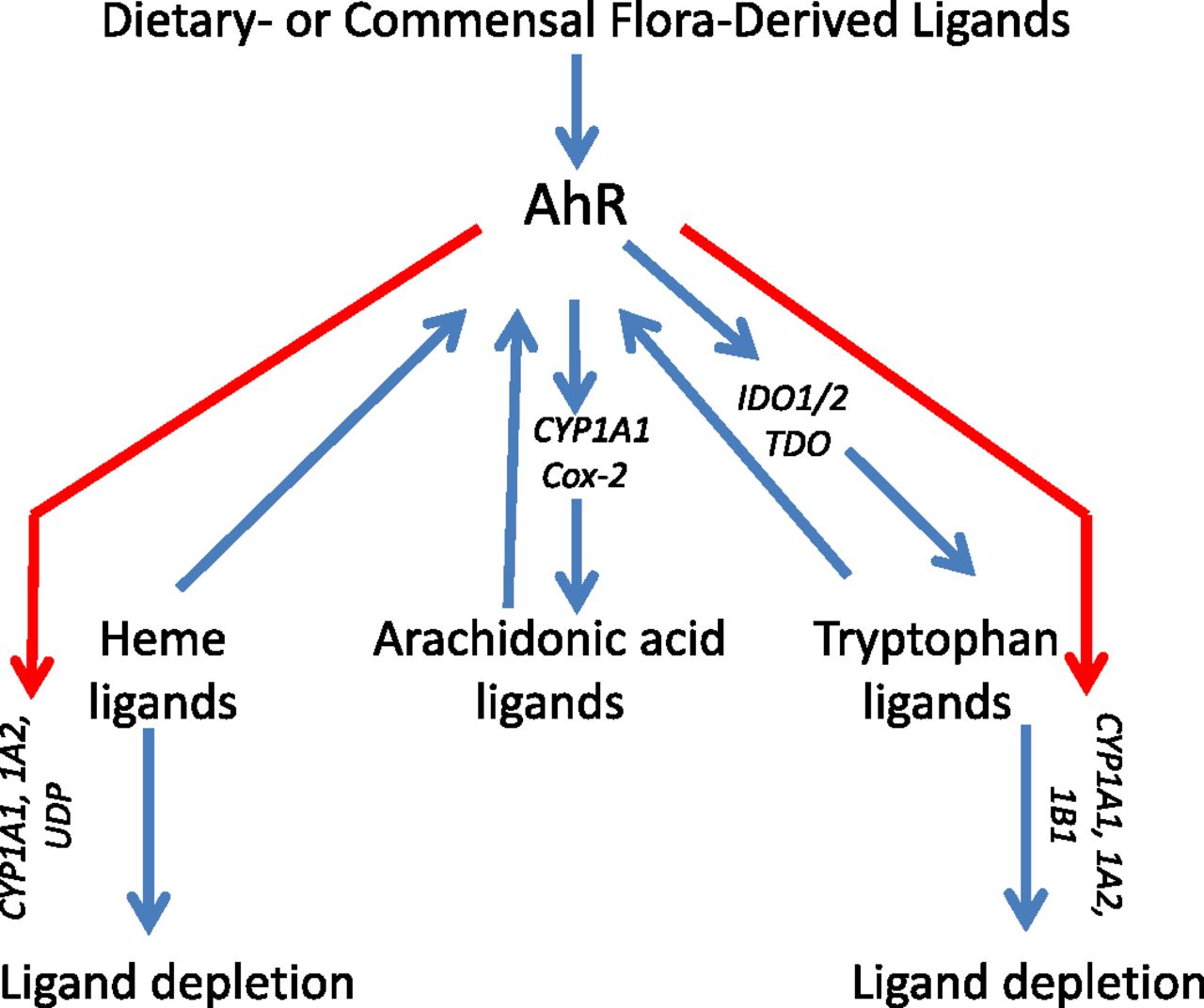
Endogenous AhR ligands and amplification of AhR activation with positive feedback loops. Exogenous ligands derived from the diet or commensal flora may initiate AhR activation. Within any given cell, heme-, arachidonic acid-, or tryptophan-derived metabolites bind to and activate the AhR. Activated AhR transcriptionally upregulates expression of genes encoding enzymes involved in arachidonic acid (CYP1A1, Cox-2) and tryptophan (CYP1A1, CYP1A2, CYP1B1) metabolism, producing AhR ligands in a localized positive feedback loop. AhR upregulation of genes encoding enzymes that degrade heme-derived [CYP1A1, CYP1A2, UDP-glucuronosyl-transferase (UDT)] and tryptophan-derived (CYP1A1, CYP1A2, CYP1B1) AhR ligands represents a negative feedback loop.
A. Indirect Evidence of Endogenous Aryl Hydrocarbon Receptor Ligands
One of the earliest results supporting the presence of endogenous AhR ligands came from studies demonstrating that CYP1A1-deficient cells exhibit a higher level of baseline AhR activity than wild-type cells (Chang and Puga, 1998). Ectopic expression of CYP1A1 decreases AhR activity to wild-type levels, suggesting that endogenous AhR ligand(s) are CYP1A1 substrates and that AhR-mediated CYP1A1 induction is, in effect, a negative feedback loop. Similarly, ectopic CYP1A2 and CYP1B1 expression decreases baseline AhR-dependent reporter activity (Chiaro et al., 2007). Additional studies using a CYP1A1-specific inhibitor, 1-(1-propynyl)pyrene, demonstrated that CYP1A1 downregulation increases AhR transcriptional activity in hepatoma cells (Levine-Fridman et al., 2004). We observed the same phenomenon after CYP1B1 knockdown in breast cancer cells (data not shown). The ability of CYP1A1 to degrade an endogenous AhR ligand was directly supported by the demonstration that CYP1A1 inhibition via ultraviolet B irradiation increases metabolism of 6-formylindolo[3,2-b]carbazole (FICZ), a postulated endogenous AhR ligand (Luecke et al., 2010; Wincent et al., 2012) (see section IV.D). Most recently, and with specific regard to the function of IL-22-secreting Th cells, it was shown that activation of the Notch signaling pathway induces production of an as yet unidentified AhR ligand capable of inducing T-cell-derived IL-22 (Alam et al., 2010). It was suggested that the source of this AhR ligand was T cells, although an increased level of AhR-inducing activity was observed when Notch signaling was induced in cocultures of T cells and APC compared with T cells alone. The identity of the putative AhR ligand(s) in these T-cell studies was not determined, although it was postulated to be a heat-labile molecule unrelated to tryptophan metabolites, previously suggested to act as endogenous AhR ligands (see section IV.D).
Perhaps the most compelling indirect evidence of endogenous, or at least “natural” AhR ligands, comes from studies with AhR−/− mice in which several developmental defects are seen including a decrease in oocyte atresia (Robles et al., 2000; Matikainen et al., 2001, 2002), ductus venosus (Lahvis et al., 2005), immature vasculature development (Lahvis et al., 2005), cardiac hypertrophy (Fernandez-Salguero et al., 1997), oculomotor defects (Chevallier et al., 2013), disproportionate red blood cell and megakaryocyte production (Lindsey and Papoutsakis, 2011), altered hematopoietic stem cell differentiation (Singh et al., 2011a), and malformation of the gut epithelial barrier (Kiss et al., 2011; Li et al., 2011). Most relevant to T-cell development and function is the failure of AhR−/− mice to generate gut-associated lymphoid leukocytes, including IL-22-secreting innate immune cells, and intraepithelial lymphocytes in both the gut and skin (Kiss et al., 2011; Li et al., 2011). Given the diversity of the effects of AhR deficiency, it seems reasonable to propose either that a single AhR ligand is pervasive throughout the developing or mature animal or, more likely, that multiple AhR ligands, potentially derived from disparate sources, act on specific tissue in a highly regulated fashion and resulting in different outcomes.
B. Heme-Derived Molecules
The finding that rats defective in UDP-glucuronosyltransferase, a key bilirubin-degrading enzyme, have elevated CYP1A1 levels (Kapitulnik and Gonzalez, 1993) hinted at the ability of this, or related heme-derived metabolites, to activate the AhR. Indeed, nanomolar concentrations of bilirubin induce CYP1A1 in mouse hepatoma cells in an AhR-dependent manner (Sinal and Bend, 1997). A related heme-degradation product, biliverdin, also activates the AhR (Phelan et al., 1998). Because the bulk of bilirubin is conjugated to albumin and thereby unavailable as an AhR ligand, its effects may be local rather than systemic. Therefore, if either biliverdin or bilirubin in fact regulates the AhR in situ, it would be expected that they do so in heme-rich organs such as liver and spleen. Interestingly, CYP1A1 and CYP1A2, along with AhR-regulated UDP-glucuronosyltransferase, contribute to bilirubin catabolism (Kapitulnik and Gonzalez, 1993; Munzel et al., 2003; Erichsen et al., 2008; Buckley and Klaassen, 2009; Bock and Kohle, 2010), suggesting a negative feedback loop consistent with that postulated by Chang and Puga (1998) and Levine-Fridman et al., 2004 (Fig. 2).
C. Arachidonic Acid Metabolites
For many years, investigators have reported an apparent correlation between outcomes seen in AhR−/− mice or after xenobiotic activation of the AhR and functions controlled by arachidonic acid metabolites, particularly inflammatory responses (Nebert and Karp, 2008). However, the exact mechanisms responsible for this association have been difficult to define, although some studies suggest that AhR activation by eicosanoids is responsible. Eicosanoids, of which over 120 have been identified, are a set of biologically activate oxygenated derivatives of arachidonic acid produced, in part, as a consequence of CYP1A1 and COX-2 enzyme activity. It is noteworthy that both the CYP1A1 and COX-2 genes are well-established AhR gene targets (Degner et al., 2007, 2009; Vogel et al., 2007), thereby linking AhR activation to prostaglandin, prostacyclin, and thromboxane synthesis. Indeed, TCDD increases expression of four dihydroxyeicosatrienoic acids and four hydroxyeicosatrienoic acids (HETEs) in an AhR-dependent manner in vivo (Bui et al., 2012). Analogous to the heme-derived AhR ligands, arachidonic acid derivatives may participate in a positive feedback loop (Fig. 2) because several prostaglandins (PGF3α, PGG2, PGH1, PGB3, PGD3, and PGH2), leukotrienes (6-trans-LTB 4, 6- trans-12-epi-LTB), dihydroxyeicosatriaenoic acids [4,5(S),6(S)-DiHETE, 5(S),6(R)-DiHETE], and at least one hydroxyeicosatrienoic acid ([12(R)-HETE]) and one lipoxin (lipoxin A4) are AhR inducers (Schaldach et al., 1999; Seidel et al., 2001; Chiaro et al., 2008a,b). Of these compounds, PGG2 and [12(R)-HETE] are the most potent and efficacious AhR activators and are capable of inducing AhR activity at nanomolar concentrations and, in some cases, inducing a 3- to 4-fold greater level of AhR-dependent reporter activity than TCDD (Seidel et al., 2001; Chiaro et al., 2008b). Nevertheless, even the weaker arachidonic acid metabolites may play a physiologic role in their respective local environments. Furthermore, the disparate molecular structure of these compounds, particularly lipoxin A4, which does not contain a ring structure, underscores the relative permissiveness of the AhR ligand-binding domain.
D. Tryptophan Metabolites
Perhaps the leading candidates for endogenous AhR ligands are tryptophan-derived metabolites. There are at least five pathways of tryptophan metabolism that have been implicated in the regulation of AhR activation:
The primary route of tryptophan metabolism in mammals is the kynurenine pathway. The most proximal metabolite, kynurenine, is produced through the activity of IDO1/2 or tryptophan-2,3-dioxygenase (Allegri et al., 2003). Kynurenine is produced by glioblastomas and regulates their survival and motility through AhR signaling (Opitz et al., 2011). More germane to autoimmunity, kynurenine induces regulatory T cells (Mezrich et al., 2010), although its source in vivo is unknown. The downstream metabolites kynurenic acid and xanthurenic acid also act as AhR ligands (Heath-Pagliuso et al., 1998; DiNatale et al., 2010). Kynurenic acid induces production of inflammatory cytokines (DiNatale et al., 2010). Importantly, IDO1 and IDO2 can be upregulated upon AhR activation (Vogel et al., 2008; Bankoti et al., 2010), again suggesting that AhR activation may result in a positive feedback loop that prolongs and/or amplifies AhR signaling in the local environment (Fig. 2).
The IDO/tryptophan-2,3-dioxygenase-independent tryptamine pathway, catalyzed by tryptophan hydroxylase and dopamine decarboxylase, also has been implicated in AhR activation. The most proximal metabolite in that pathway, tryptamine, is a potent AhR activator and may be both a direct AhR ligand (Heath-Pagliuso et al., 1998) and a precursor of a downstream AhR ligand(s) (Vikstrom Bergander et al., 2012). Indeed, at least one metabolite downstream of tryptamine, indole acetic acid, is an AhR ligand (Heath-Pagliuso et al., 1998). Of note, tryptamine is a competitive CYP1A1 substrate, a result consistent with the prediction that endogenous AhR ligand(s) is degraded by CYP1 enzymes (Chiaro et al., 2007).
The serotonin pathway may be directly or indirectly connected to AhR activation. 5-Hydroxytryptophan, a proximal serotonin metabolite, is an AhR agonist of modest potency (Bittinger et al., 2003). In our hands, serotonin itself is a weak AhR activator in an AhR-dependent reporter assay (data not shown). Perhaps more important to AhR regulation is the metabolic pathway distal to serotonin. In one branch of the serotonin pathway represented by tryptophan → 5-hydroy-l-tryptophan → serotonin → N-acetyl-serotonin → melatonin → 6-hydroxymelatonin, melatonin conversion is catalyzed by CYP1A1, CYP1A2, and CYP1B1. Therefore, upregulation of CYP1 enzymes could result in a metabolic sink that depletes tryptophan stores and reduces production of endogenous AhR ligands in the kynurenine and/or tryptamine pathways (Fig. 2). Consequently, an increase in AhR activity after exposure to a given compound (Vikstrom Bergander et al., 2012) could be mistaken for evidence that the compound is an AhR ligand when in reality the increase in AhR activity could be because of blocking of the serotonin pathway metabolic sink and the subsequent increase in concentration of AhR ligands from the kynurenine, tryptamine, or tryptophan photometabolite (see below) pathways.
It was first suggested in 1987 that tryptophan photometabolites represent endogenous AhR ligands (Rannug et al., 1987). Since then, many studies have demonstrated that 6-formylindolo[3,2-b]carbazole (FICZ) binds to the AhR with high affinity and initiates AhR signaling at nanomolar concentrations (Rannug et al., 1987; Wei et al., 1999, 2000; Oberg et al., 2005; Goryo et al., 2007; Mukai and Tischkau, 2007; Wincent et al., 2009, 2012). FICZ can contribute to (Kostyuk et al., 2012) or block (Jeong et al., 2012) inflammatory responses in a tissue- and context-specific fashion. FICZ also influences responses to ultraviolet light, genomic stability, and circadian rhythms, all postulated to be AhR mediated (Wincent et al., 2012). At least one additional tryptophan photometabolite, 1-(1H-indol-3-yl)-9H-pyrido[3,4-b]indole, has been identified and shown to exhibit a similar potency and efficacy as FICZ (Diani-Moore et al., 2011). Although initially thought to exert only local AhR-mediated effects in the skin, the influence of these photometabolites may be systemic as FICZ has been found in human urine (Wincent et al., 2009). Finally, FICZ is readily metabolized by CYP1A1, CYP1A2, and CYP1B1 (Wincent et al., 2012), satisfying a proposed criterion for endogenous AhR ligands (Diani-Moore et al., 2011).
Plant-derived phytochemicals were among the earliest of postulated “natural” AhR ligands. Plant-derived indirubin and indigo are AhR ligands of relatively high potency (Adachi et al., 2001). However, the extremely low concentration in which they are found in human urine (∼0.2 nM) and the failure to detect either compound in mammalian tissue suggests that they are not meaningful AhR inducers under normal physiologic conditions. Indole-3-carbinol and its condensation derivatives 3,3′-diidolmethane, indolo[3,2-b]carbazole, and 2-(indol-3-ylmethyl)-3,3′-diindolylmethane, are derived from cruciferous vegetables and exhibit strong affinity for the AhR (Bjeldanes et al., 1991; Chen et al., 1996, 1998; Safe et al., 1999; Safe and McDougal, 2002; Ociepa-Zawal et al., 2007; Degner et al., 2009). These or other phytochemicals are very likely to be important in the context of T-cell development, autoimmunity, and inflammation inasmuch as dietary AhR ligands are critical for the development of gut-associated intraepithelial lymphocytes and maintenance of gut epithelial barrier integrity (Kiss et al., 2011; Li et al., 2011). Furthermore, the involvement of yet more tryptophan/indole derivatives in AhR activation suggests another intriguing possibility that indole, produced from tryptophan by a variety of commensal gut microflora, serves as the precursor for the generation of one or more “endogenous” AhR ligands. If confirmed, this would directly tie the microbiome to production of AhR ligands capable of influencing T-cell development and function at multiple levels.
E. Aryl Hydrocarbon Receptor Feedback Loops
From the results described above, it is apparent that control of the production and degradation of endogenous AhR ligands is complex (Fig. 2). The likelihood of positive feedback loops could significantly amplify the signal induced even by weak AhR ligands. Their existence also suggests the possibility that, once homeostasis is perturbed by exposure to low levels of environmental AhR ligands or AhR-targeted therapeutics, a self-perpetuating loop of AhR signaling may persist, even if the initiating ligand is metabolized and cleared. Although this may be undesirable in the context of environmental chemical exposures, it may be useful for therapeutic strategies aimed at activating the AhR. The fact that the AhR induces enzymes that degrade some endogenous AhR ligands also adds a level of complexity and unpredictability. Compounds that bind to and activate the AhR also tend to be substrates of AhR-regulated CYP1 enzymes, e.g., tryptamine (Heath-Pagliuso et al., 1998), FICZ (Wei et al., 2000), and indole-3-carbinol derivatives (Bittinger et al., 2003). Indeed, it has been suggested that some compounds that induce AhR signaling do so not because they are AhR ligands but because they reduce catabolism of bona fide endogenous ligands (Wincent et al., 2012). Consequently, the magnitude and persistence of AhR signaling is a function of the affinity of a compound for the AhR, its activity as a CYP1 substrate (or inhibitor), the AhR-independent factors that control CYP1 levels in any given cell type or tissue, and the propensity for the AhR in any given tissue to amplify expression of enzymes involved in endogenous ligand synthesis. Therefore, predictions of what any particular AhR ligand may do in a given context remain difficult to make. Therefore, empirical testing may be the only way to determine if any given AhR ligand, e.g., an AhR-targeted therapeutic, induces any AhR-mediated toxicity.
V. Aryl Hydrocarbon Receptor as a Therapeutic Target
Because of its effects on the regulatory T-cell compartment, the AhR is considered a potential therapeutic target for the treatment of autoimmune disorders. Indeed, the administration of certain AhR ligands, including TCDD, the quintessential environmental AhR ligand, leads to the expansion of functional Tregs that arrest the development of experimental autoimmune encephalomyelitis (EAE) (Quintana et al., 2008), experimental autoimmune uveoretinitis (Zhang et al., 2009), colitis (Benson and Shepherd, 2011; Singh et al., 2011b), and spontaneous autoimmune diabetes (Kerkvliet et al., 2009). Some limitations, however, will have to be overcome to translate these findings to the treatment of human autoimmune disorders.
One of these limitations is the broad tissue expression pattern of the AhR, potentially making it difficult to selectively target specific tissues after systemic administration of AhR modulators. Of particular concern for autoimmunity is the observation that development of both inflammatory Th17 cells, which promote organ-specific autoimmunity, and Treg cells, which oppose autoimmunity, are facilitated by AhR activation. Thus, systemically administered AhR ligands could, under different circumstances, exacerbate or ameliorate autoimmunity. To overcome this uncertainty, we have constructed nanoparticles (NPs) with the goal of delivering AhR ligands directly to APCs (Fig. 3). Nanotechnology offers new opportunities for therapeutic intervention in immune-mediated diseases. NP-based drug delivery systems increase the half-life of compounds, facilitating the delivery of one or several compounds to specific cell-types in vivo (Sanvicens and Marco, 2008). Based on the tolerogenic effects of AhR activation in DCs, we engineered NPs to co-deliver the AhR ligand 2-(1H-indol-3-ylcarbonyl)-4-thiazolecarboxylic acid methyl ester (ITE), which induces a tolerogenic phenotype in DCs in vitro, together with tissue-specific antigens (Yeste et al., 2012).
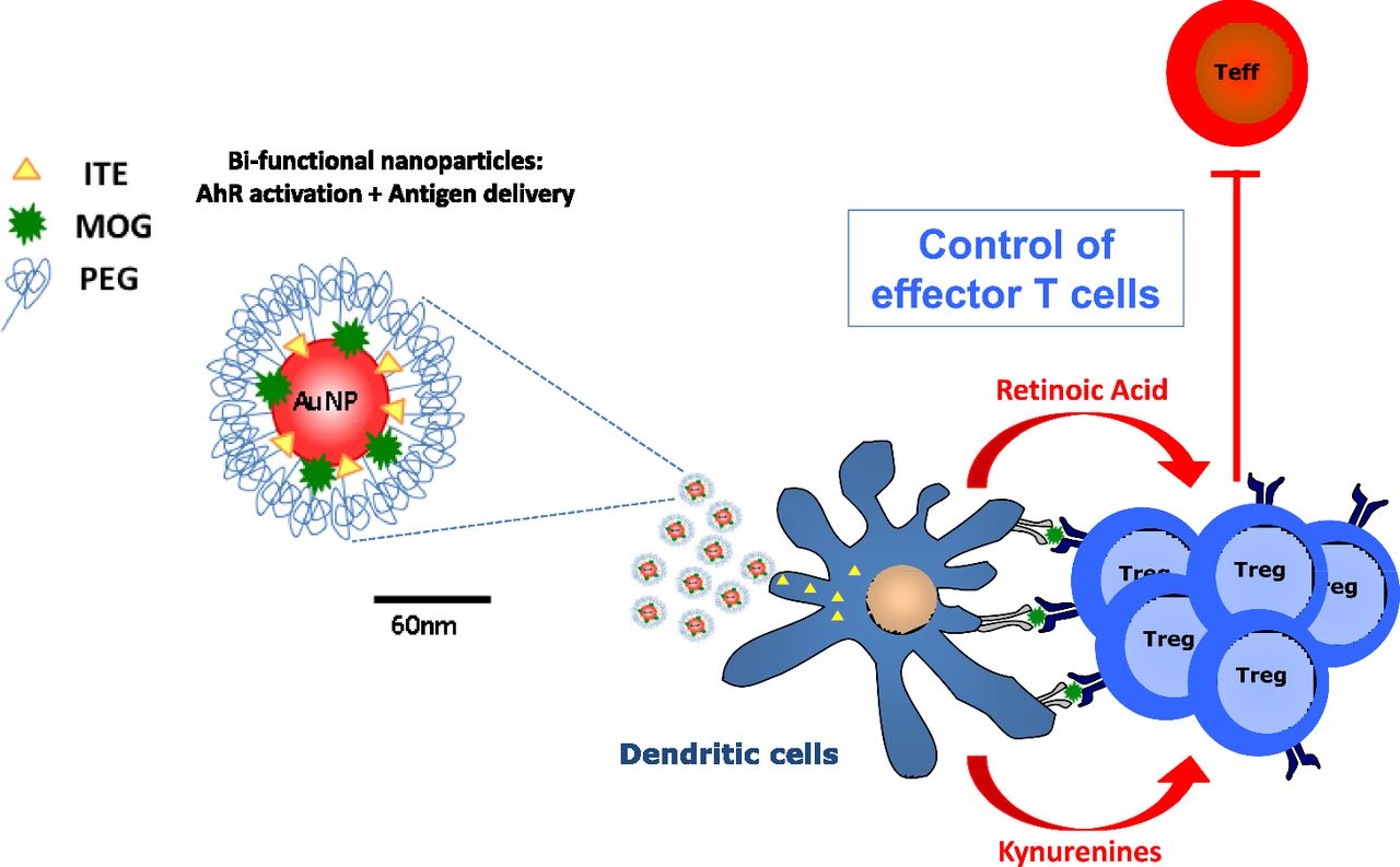
Bifunctional nanoparticles for the induction of antigen-specific immune tolerance. Gold nanoparticles coated with polyethylene glycol (PEG) are engineered ligand ITE and the central nervous system antigen myelin oligodendrocyte protein (MOG) to dendritic cells in vivo. Dendritic cells that produce kynurenines (postulated tryptophan-derived endogenous AhR ligands) and retinoic acid induce and/or expand MOG-specific regulatory T cells (Treg) through retinoic acid receptor- and AhR-dependent signaling pathways. Treg cells actively suppress effector T-cell function (Teff), thereby inhibiting central nervous system autoimmunity.
NPs protected ITE from degradation by liver enzymes, increasing its half-life in vivo (Yeste et al., 2012). After intravenous administration, ITE-loaded NPs activated AhR signaling in DCs in vivo. NP-triggered AhR activation in DCs induces a tolerogenic phenotype characterized by the reduced ability to generate Th1 and Th17 cells, concomitant with an increased ability to promote FoxP3+ Treg differentiation. Accordingly, NPs loaded with ITE and myelin antigens expand central nervous system-specific FoxP3+ Tregs that suppress EAE, both in preventive and therapeutic paradigms. Taken together, these data suggest that NP-based administration offers a new avenue for the selective activation of AhR in specific cell populations for therapeutic purposes.
A second issue that must be addressed when considering the use of AhR agonists for treatment of autoimmune disorders, or other diseases for that matter, is our lack of understanding of how different AhR ligands induce different outcomes. For example, in the same animal model of EAE, TCDD, or ITE treatment induces Treg cells and suppresses autoimmunity, whereas another ligand, FICZ, produces the opposite effect, i.e., it induces Th17 cells and exacerbates disease (Quintana et al., 2008). Notably, both ITE and FICZ may be physiologically relevant endogenous AhR ligands (Song et al., 2002; Wincent et al., 2009), suggesting the possibility that different ligands are generated in situ in a context-specific manner to induce different AhR-dependent outcomes.
In the field of toxicology, the ability of different AhR ligands to induce different biologic outcomes has been well established. Most frequently, these differences have been attributed to differences in ligand persistence, a function of ligand metabolism. For example, 7.12-dimethylbenz[a]anthracene is readily metabolized by AhR-induced monooxygenases (CYP1A1, CYP1A2, CYP1B1) and induces significant bone marrow toxicity in vivo and in vitro through apoptosis induction (Yamaguchi et al., 1997; Mann et al., 1999; Allan et al., 2003; Ryu et al., 2005; Teague et al., 2010). TCDD, which is not readily metabolized and, consequently, persists in vivo for 7–10 years, does not induce apoptosis in the bone marrow. Differences in biologic outcomes with different AhR ligands also have been attributed to different ligand affinities for the AhR. However, it is unlikely that affinity differences alone account for the opposite effects of, for example, TCDD and FICZ, because both of these compounds bind the AhR with comparable affinities (Kd = 0.07–1.7 nM) (Furuhashi et al., 1986; Wei et al., 2000; Nguyen and Bradfield, 2008).
This variability in biologic outcomes with different AhR ligands has another important implication, i.e., not all AhR ligands are toxic and/or carcinogenic. For example, although readily metabolized polycyclic aromatic hydrocarbons, such as benzo[a]pyrene, and poorly metabolized TCDD are both toxic and carcinogenic, there is no evidence to suggest that any of the “natural” AhR ligands, including FICZ, ITE, eicosanoids, or kynurenine pathway metabolites, all of which induce CYP1 enzymes, are either toxic or carcinogenic. Therefore, an AhR-targeted therapeutic need not invoke the specter of unacceptable toxic side effects.
VI. Conclusions
AhR modulates the activity of the immune system in response to structurally heterogeneous ligands of diverse origin. These features of AhR are reminiscent of the effects on immunity of Toll-like receptor (TLR) signaling triggered by self and non-self agonists (Kawai and Akira, 2010). For example, both AhR and TLRs are evolutionary conserved systems that control innate and adaptive immunity in response to diverse environmental and endogenous cues. Indeed, AhR and TLR signaling intersect and cross-regulate each other. TLR activation upregulates AhR expression (Amit et al., 2009; Nguyen et al., 2010a), and AhR acts as a negative regulator of TLR signaling (Kimura et al., 2009; Mezrich et al., 2010; Nguyen et al., 2010; Quintana et al., 2010b). Thus, AhR, TLRs, and additional signaling pathways cross-talk to adjust the activity of the immune system in response to signals provided by the environment, the diet, and the commensal flora. A corollary to this interpretation is that molecular targeting of the AhR, like molecular targeting of TLRs, provides an opportunity for therapeutic modulation of the immune response.
Several ligands operate in vivo, probably in a tissue-specific manner, to activate the AhR. The specific combination of the cytokine milieu, the specific tissue and ligand involved, determines whether the activation of AhR results in the promotion or the suppression of a specific immune response. Thus, controlled therapeutic targeting of the AhR requires the identification of ligands that promote the desired immune response without unwanted toxic or carcinogenic effects. Immunomodulatory endogenous ligands that lack toxicity and carcinogenicity have already been identified, suggesting that it is possible to design AhR-targeting compounds for the therapeutic modulation of the immune response. The half-life and therapeutic efficacy of these nontoxic AhR ligands might be increased with the use of nanomaterials for the targeted activation of AhR in specific cell types.
In conclusion, the AhR controls important immune processes in response to endogenous and environmental cues. Consequently, the AhR and its signaling pathway offer plausible molecular mechanisms through which environmental and endogenous ligands may control immunity and autoimmunity while providing us with new opportunities for targeted, therapeutic modulation of the immune response.
Acknowledgments
The authors want to thank the members of their laboratories for valuable discussions.
Authorship Contributions
Wrote or contributed to the writing of the manuscript: Quintana and Sherr.
Footnotes
Research in the Quintana laboratory was supported by the National Institutes of Health National Institute of Allergy and Infectious Diseases [Grants AI075285 and AI093903]; the National Multiple Sclerosis Society; and the Juvenile Diabetes Research Foundation. Research in the Sherr laboratory was supported by the National Institutes of Health National Institute of Environmental Health Sciences [Grants P01ES11624 and P42ES007381]; and the Art beCAUSE Breast Cancer Foundation.
Abbreviations
- AhR
- aryl hydrocarbon receptor
- APCs
- antigen presenting cells
- DCs
- dendritic cells
- EAE
- experimental autoimmune encephalomyelitis
- FICZ
- 6-formylindolo[3,2-b]carbazole
- HETEs
- 4-hydroxyeicosatrienoic acids
- HSP90
- 90-kDa heat shock protein
- IDO
- indoleamine 2,3-dioxygenase
- IL
- interleukin
- ITE
- 2-(1H-indol-3-ylcarbonyl)-4-thiazolecarboxylic acid methyl ester
- iTregs
- induced FoxP3+ Tregs generated in the periphery
- NPs
- nanoparticles
- nTregs
- natural FoxP3+ Tregs generated in the thymus
- PG
- prostaglandin
- RA
- retinoic acid
- TCDD
- 2,3,7,8-tetracholrodibenzo-p-dioxin
- TLR
- Toll-like receptor
- Tr1 cells
- Type 1 regulatory T cells
- Tregs
- regulatory T cells
- Copyright © 2013 by The American Society for Pharmacology and Experimental Therapeutics
References
In this issue


Jump to section
- Article
- Abstract
- I. Introduction
- II. Aryl Hydrocarbon Receptor Signaling Pathways
- III. The Aryl Hydrocarbon Receptor in Immunity and Autoimmunity
- IV. Endogenous Aryl Hydrocarbon Receptor Ligands and Amplification of Aryl Hydrocarbon Receptor Activation with Positive Feedback Loops
- V. Aryl Hydrocarbon Receptor as a Therapeutic Target
- VI. Conclusions
- Acknowledgments
- Authorship Contributions
- Footnotes
- Abbreviations
- References
- Figures & Data
- Info & Metrics
- eLetters